Dissection of thorax from a posterior approach
Deep musculature.
Stanford holds the copyright to the David L. Bassett anatomical images and has assigned
Creative Commons license Attribution-Share
Alike 4.0 International to all of the images.
For additional information regarding use and permissions,
please contact the Medical History Center.
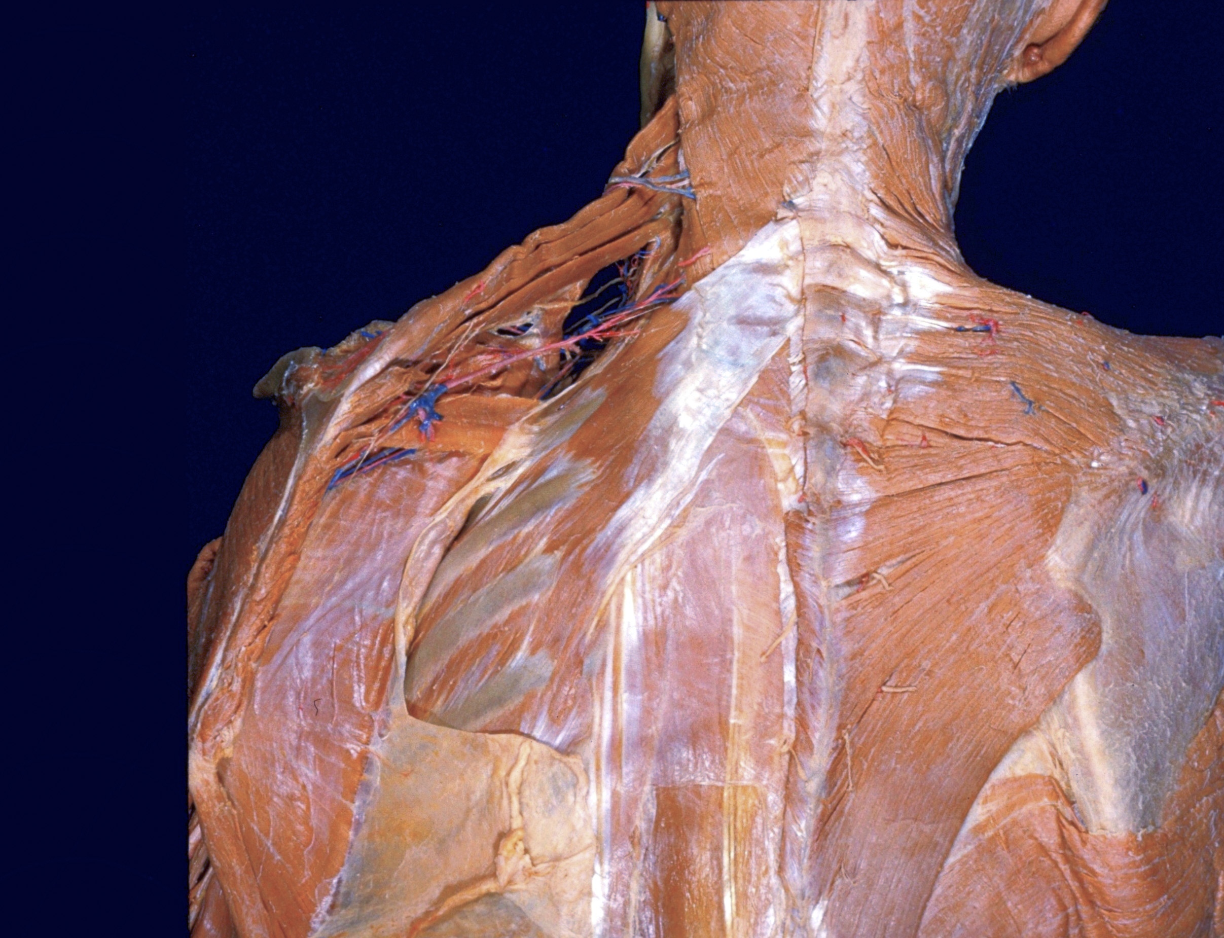
Image #131-1
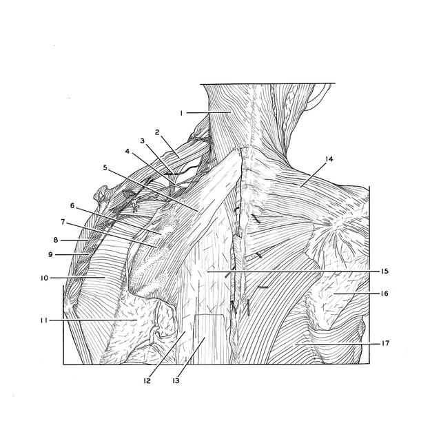
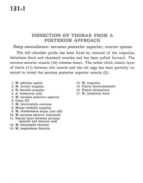
Dissection of thorax from a posterior approach
Deep musculature.
The left shoulder girdle has been freed by removal of the trapezius, latissimus dorsi and rhomboid muscles and has been pulled forward. The serratus anterior muscle (10) remains intact. The rather thick, elastic layer of fascia (11) between this muscle and the rib cage has been partially resected to reveal the serratus posterior superior muscle (5).
- Splenius capitis muscle
- Levator scapulae muscle
- Dorsal scapular nerve
- Transverse colli artery
- Serratus posterior superior muscle
- Rib III
- External intercostal muscle
- Medial scapular margin
- Rhomboid major muscle (cut off)
- Serratus anterior muscle (elevated)
- Fascial layer between serratus anterior and thoracic wall
- Iliocostalis thoracis muscle
- Longissimus thoracis muscle
- Trapezius muscle
- Thoracolumbar fascia
- Fascia infraspinata
- Latissimus dorsi muscle