Dissection of diaphragm
Diaphragm viewed from above, pleura removed
Stanford holds the copyright to the David L. Bassett anatomical images and has assigned
Creative Commons license Attribution-Share
Alike 4.0 International to all of the images.
For additional information regarding use and permissions,
please contact the Medical History Center.
Image #130-1
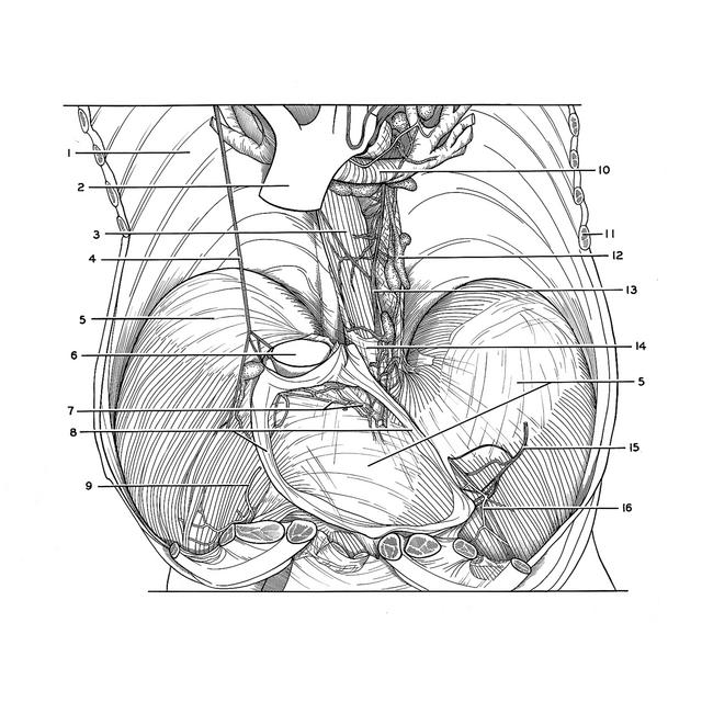
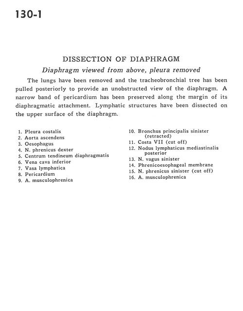
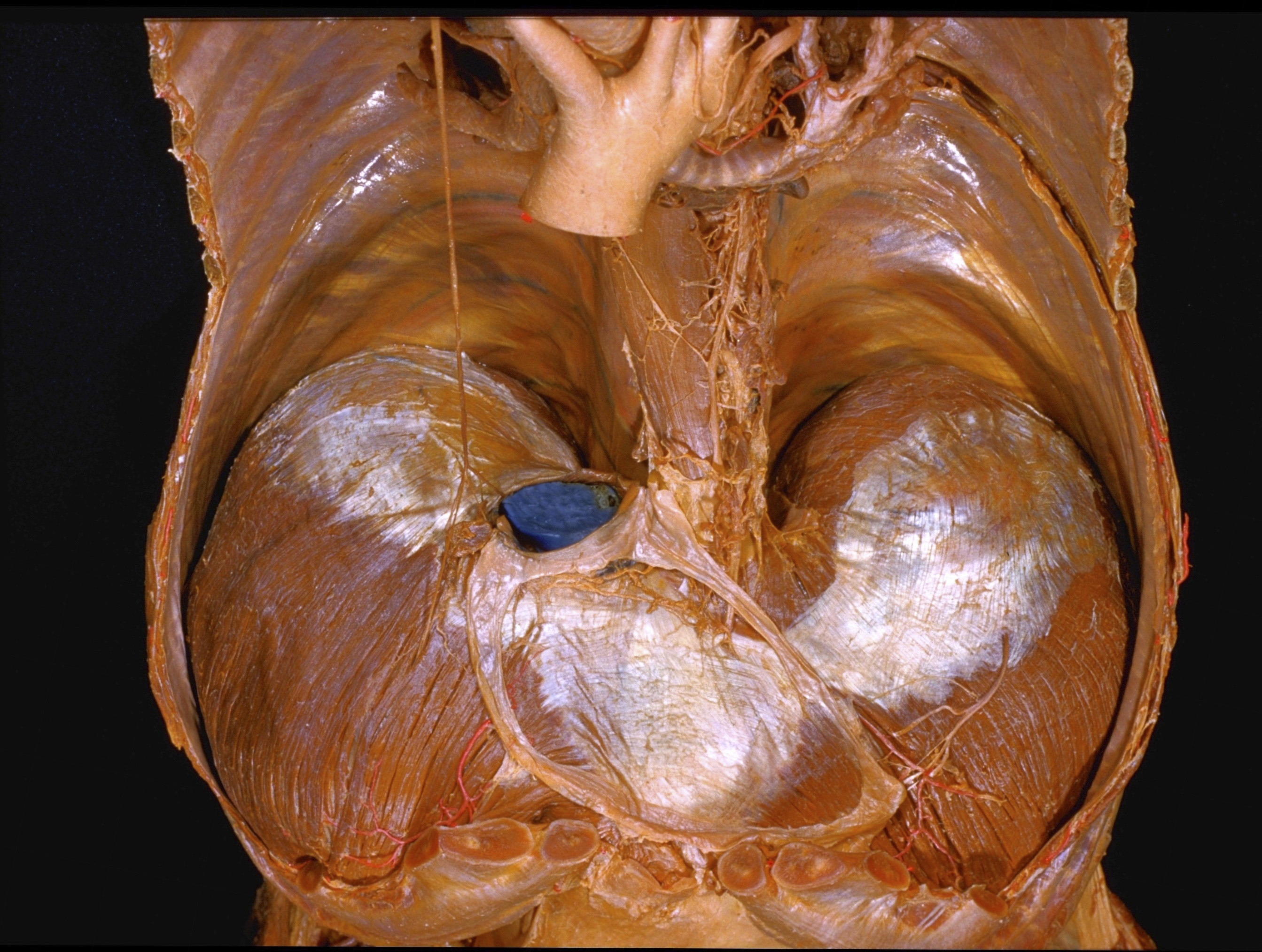
Dissection of diaphragm
Diaphragm viewed from above, pleura removed
The lungs have been removed and the tracheobronchial tree has been pulled posteriorly to provide an unobstructed view of the diaphragm. A narrow band of pericardium has been preserved along the margin of its diaphragmatic attachment. Lymphatic structures have been dissected on the upper surface of the diaphragm.
- Costal pleura
- Ascending aorta
- Esophagus
- Phrenic nerve right
- Central tendon of diaphragm
- Inferior vena cava
- Lymph vessel
- Pericardium
- Musculophrenic artery
- Left main bronchus (retracted)
- Rib VII (cut off)
- Posterior mediastinal lymph node
- Left vagus nerve
- Phrenicoesophageal membrane
- Phrenic nerve left (cut off)
- Musculophrenic artery