Dissection of mediastinum and paravertebral structures
Superior mediastinum.
Stanford holds the copyright to the David L. Bassett anatomical images and has assigned
Creative Commons license Attribution-Share
Alike 4.0 International to all of the images.
For additional information regarding use and permissions,
please contact the Medical History Center.
Image #127-2
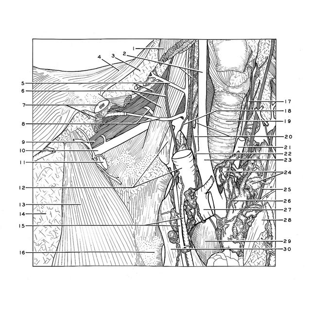
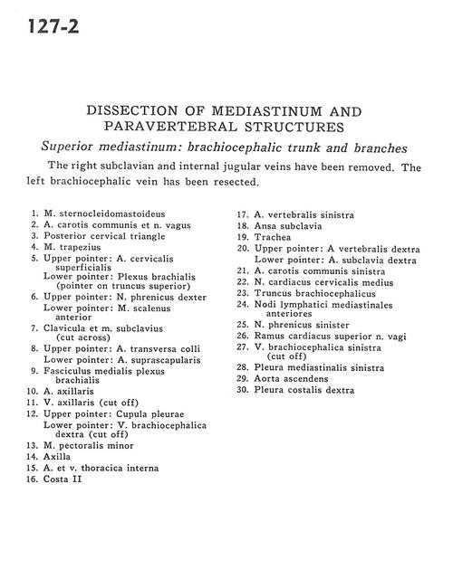
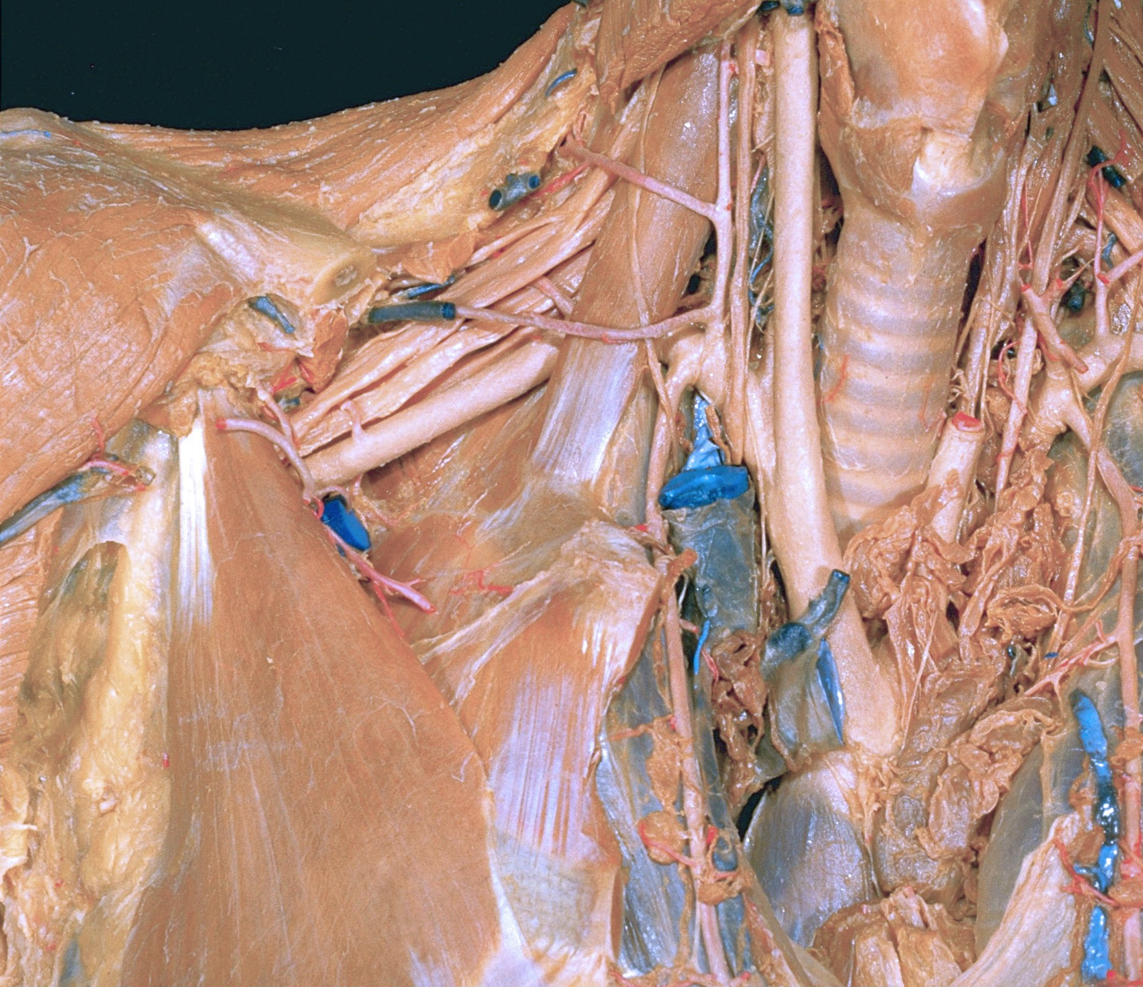
Dissection of mediastinum and paravertebral structures
Superior mediastinum.
The right subclavian and internal jugular veins have been removed. The left brachiocephalic vein has been resected.
- Sternocleidomastoid muscle
- Common carotid artery and vagus nerve
- Posterior cervical triangle
- Trapezius muscle
- Upper pointer: Superficial cervical artery Lower pointer: Brachial plexus (pointer on superior trunk)
- Upper pointer: Right phrenic nerve Lower pointer: Anterior scalene muscle
- Clavicle and subclavius muscle (cut across)
- Upper pointer: Transverse colli artery Lower pointer: Suprascapular artery
- Medial cord brachial plexus
- Axillary artery
- Axillary vein (cut off)
- Upper pointer: Cupula pleurae Lower pointer: Right brachiocephalic vein (cut oft)
- Pectoralis minor muscle
- Axilla
- Internal thoracic artery and vein
- Rib II
- Left vertebral artery
- Ansa subclavia
- Trachea
- Upper pointer: Right vertebral artery Lower pointer: Right subclavian artery
- Left common carotid artery
- Middle cervical cardiac nerve
- Brachiocephalic trunk
- Anterior mediastinal lymph nodes
- Left phrenic nerve
- Superior cardiac branch vagus nerve
- Left brachiocephalic vein (cut off)
- Left mediastinal pleura
- Ascending aorta
- Right costal pleura