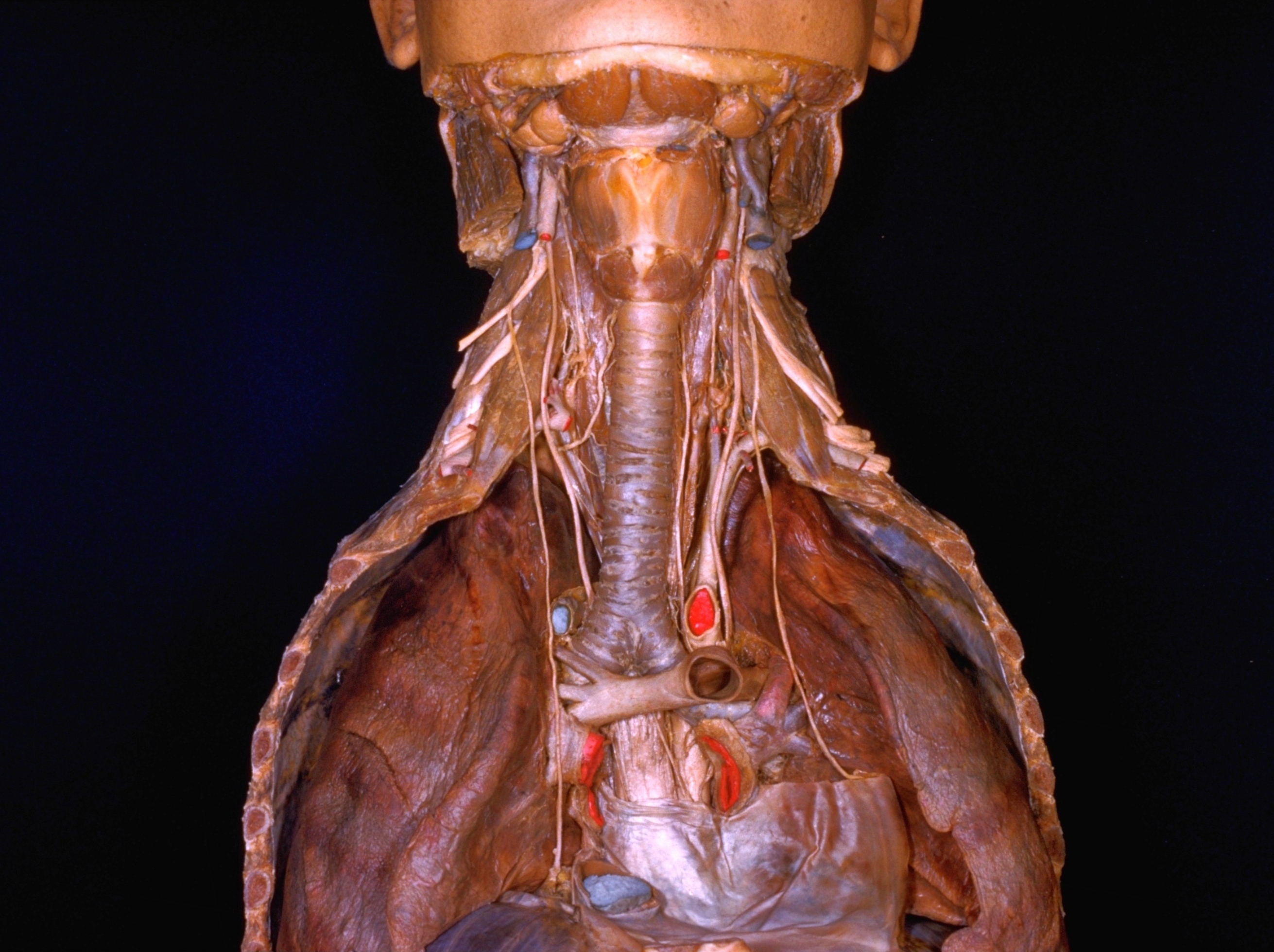
Dissection of lungs in situ
Larynx, trachea, main bronchi and lungs
Stanford holds the copyright to the David L. Bassett anatomical images and has assigned
Creative Commons license Attribution-Share
Alike 4.0 International to all of the images.
For additional information regarding use and permissions,
please contact the Medical History Center.
Image #123-5
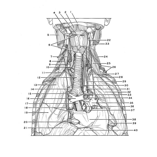
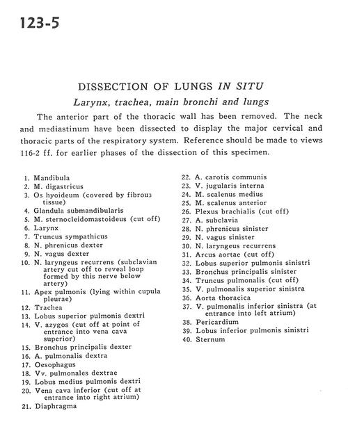
Dissection of lungs in situ
Larynx, trachea, main bronchi and lungs
The anterior part of the thoracic wall has been removed. The neck and mediastinum have been dissected to display the major cervical and thoracic parts of the respiratory system. Reference should be made to views 116-ff. for earlier phases of the dissection of this specimen.
- Mandible
- Digastric muscle
- Hyoid bone (covered by fibrous tissue)
- Submandibular gland
- Sternocleidomastoid muscle (cut off)
- Larynx
- Sympathetic trunk
- Phrenic nerve right
- Right vagus nerve
- Recurrent laryngeal nerve (subclavian artery cut off to reveal loop formed by this nerve below artery)
- Apex of lung (lying within cupula pleurae)
- Trachea
- Upper lobe right lung
- Azygos vein (cut off at point of entrance into superior vena cava)
- Right main bronchus
- Right pulmonary artery
- Esophagus
- Right pulmonary veins
- Middle lobe right lung
- Inferior vena cava (cut off at entrance into right atrium)
- Diaphragm
- Common carotid artery
- Internal jugular vein
- Middle scalene muscle
- Anterior scalene muscle
- Brachial plexus (cut off)
- Subclavian artery
- Phrenic nerve left
- Vagus nerve left
- Recurrent laryngeal nerve
- Aortic arch (cut off)
- Upper lobe left lung
- Left main bronchus
- Pulmonary trunk (cut off)
- Left superior pulmonary vein
- Thoracic aorta
- Left inferior pulmonary vein (at entrance into left atrium)
- Pericardium
- Lower lobe left lung
- Sternum