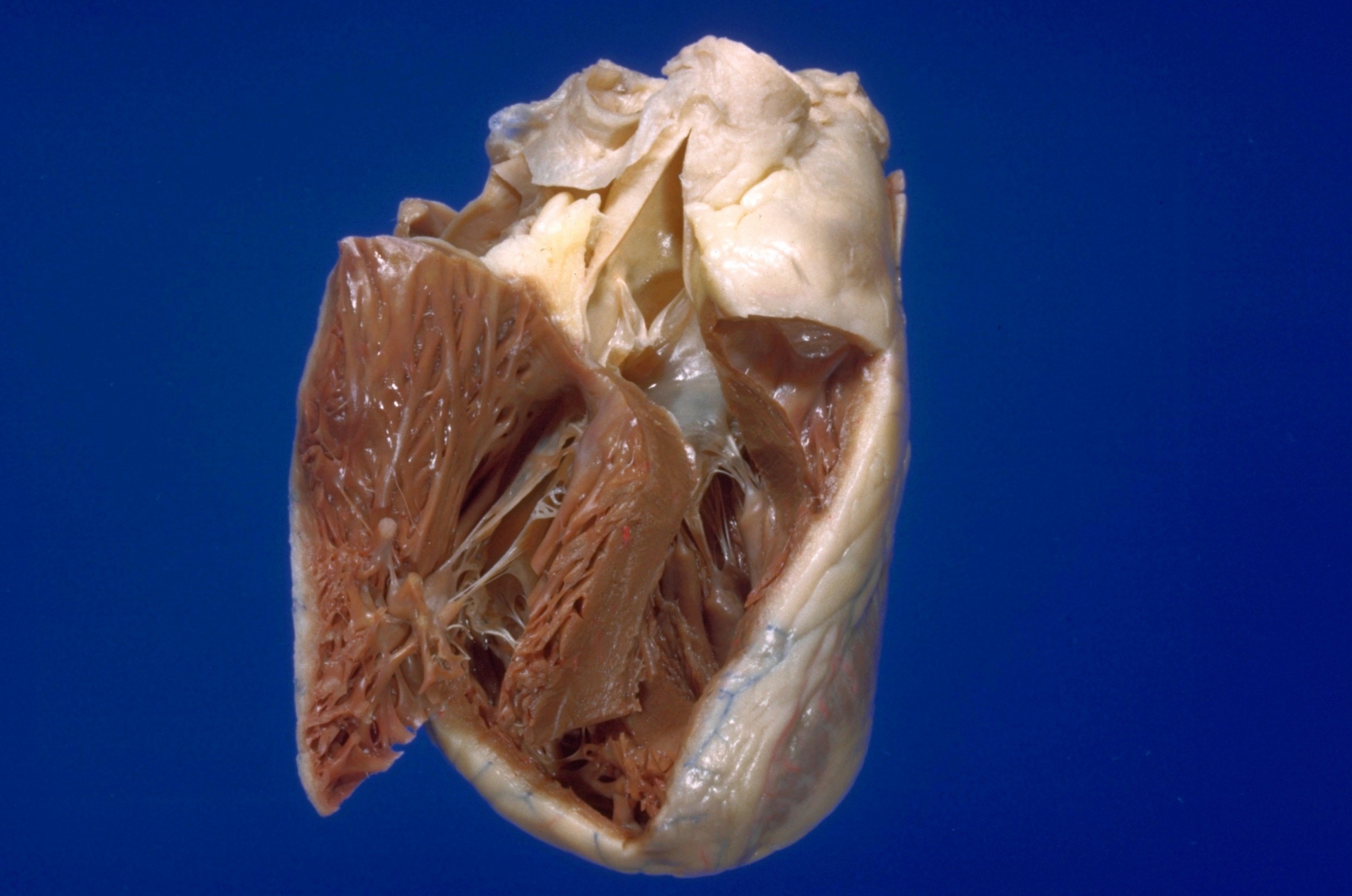
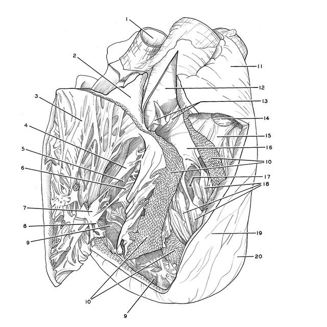
Detailed dissection of heart
Interior of right and left ventricles, anterior view
Stanford holds the copyright to the David L. Bassett anatomical images and has assigned
Creative Commons license Attribution-Share
Alike 4.0 International to all of the images.
For additional information regarding use and permissions,
please contact the Medical History Center.
Image #122-4
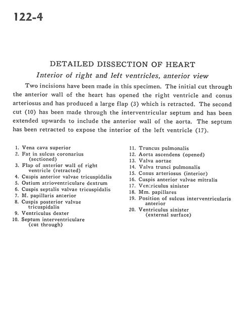
Detailed dissection of heart
Interior of right and left ventricles, anterior view
Two incisions have been made in this specimen. The initial cut through the anterior wall of the heart has opened the right ventricle and conus arteriosus and has produced a large flap (3) which is retracted. The second cut (10) has been made through the interventricular septum and has been extended upwards to include the anterior wall of the aorta. The septum has been retracted to expose the interior of the left ventricle (17).
- Superior vena cava
- Fat in coronary sulcus (sectioned)
- Flap of anterior wall of right ventricle (retracted)
- Anterior cusp tricuspid valve
- Right atrioventricular opening
- Septal (medial) cusp of tricuspid valve
- Anterior papillary muscle
- Posterior cusp tricuspid valve
- Right ventricle
- Interventricular septum (cut through)
- Pulmonary trunk
- Ascending aorta (opened)
- Aortic valve
- Valve of pulmonary trunk
- Conus arteriosus (interior)
- Anterior cusp of mitral valve
- Left ventricle left
- Papillary muscles
- Position of anterior interventricular sulcus
- Left ventricle (external surface)