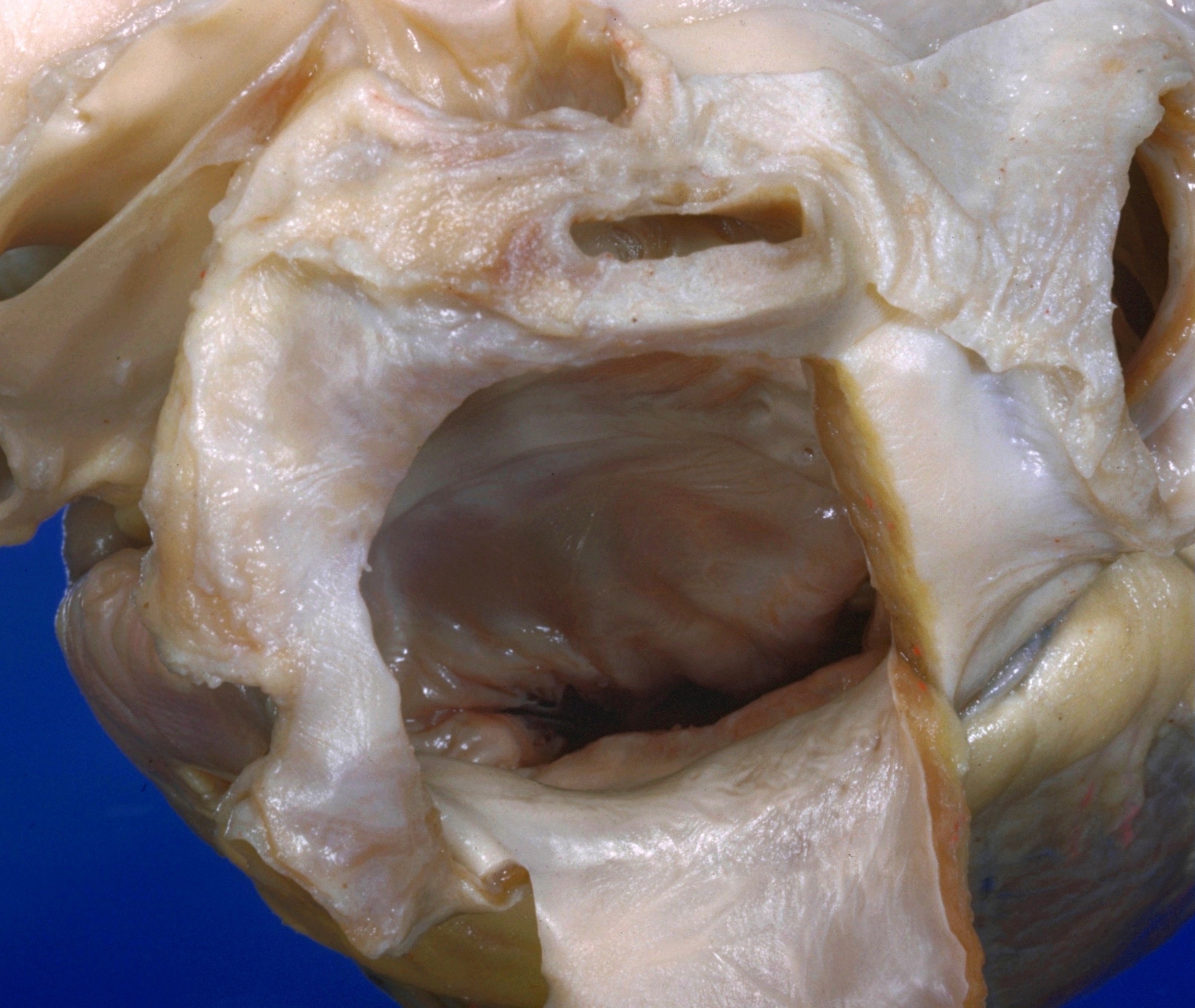
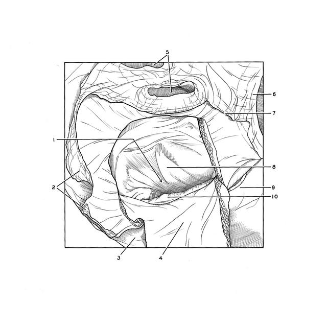
Detailed dissection of heart
Mitral valve viewed through left atrium
Stanford holds the copyright to the David L. Bassett anatomical images and has assigned
Creative Commons license Attribution-Share
Alike 4.0 International to all of the images.
For additional information regarding use and permissions,
please contact the Medical History Center.
Image #122-2
Detailed dissection of heart
Mitral valve viewed through left atrium
The specimen shown in the previous view has been turned so that the view is directed downward through the ostium of the mitral valve (1).
- Left atrioventricular opening
- Left pulmonary veins (upper vein obscured)
- Fat in posterior part of coronary sulcus
- Reflected flap of posterior atrial wall
- Right pulmonary veins
- Inferior vena cava (at point of entrance into right atrium)
- Line of reflection of serous pericardium
- Anterior cusp of mitral valve
- Fat in coronary sulcus overlying coronary sinus
- Posterior cusp of mitral valve