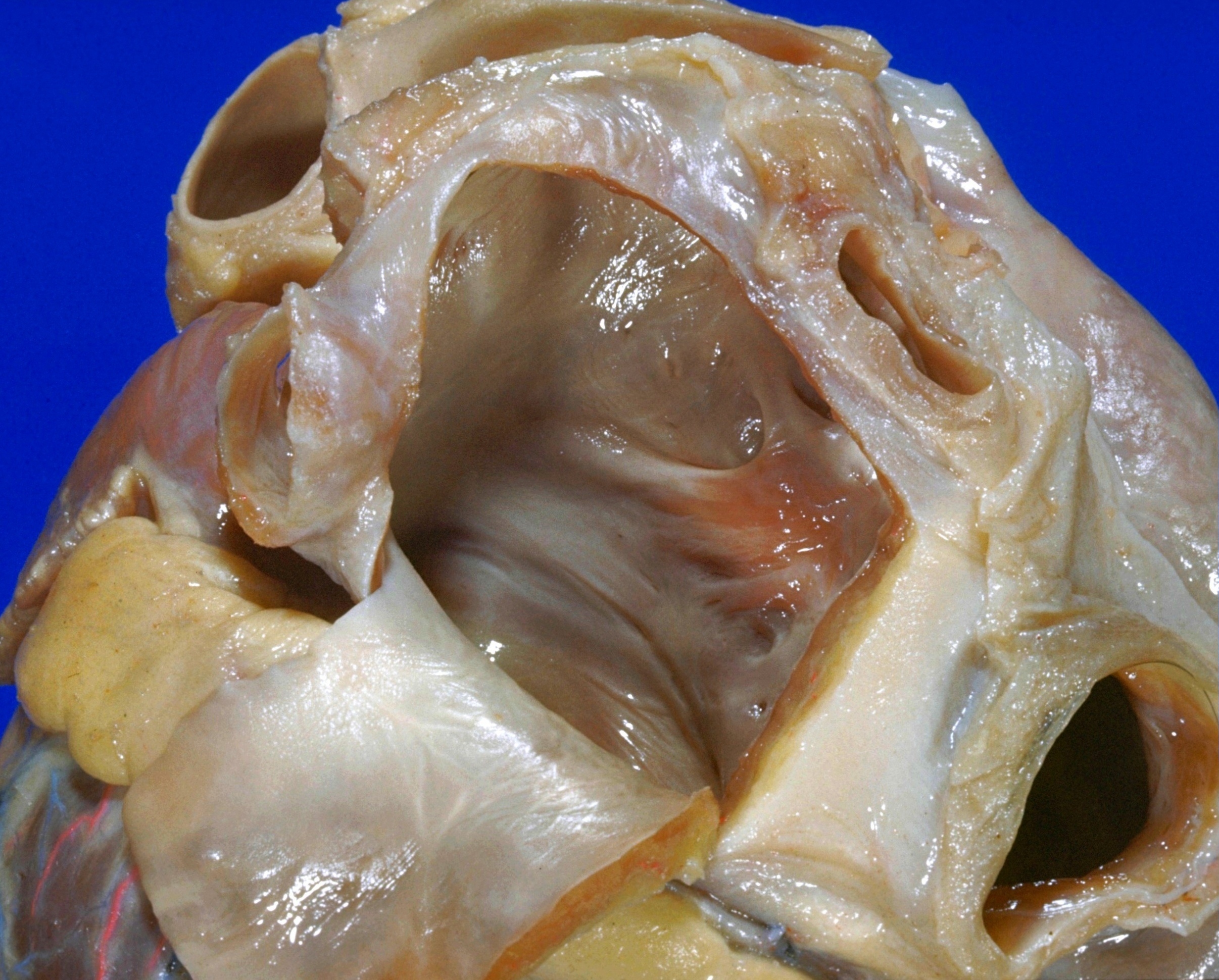
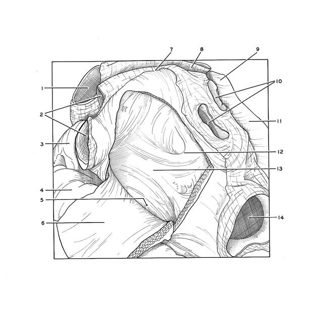
Detailed dissection of heart
Interior of left atrium, posterior view
Stanford holds the copyright to the David L. Bassett anatomical images and has assigned
Creative Commons license Attribution-Share
Alike 4.0 International to all of the images.
For additional information regarding use and permissions,
please contact the Medical History Center.
Image #122-1
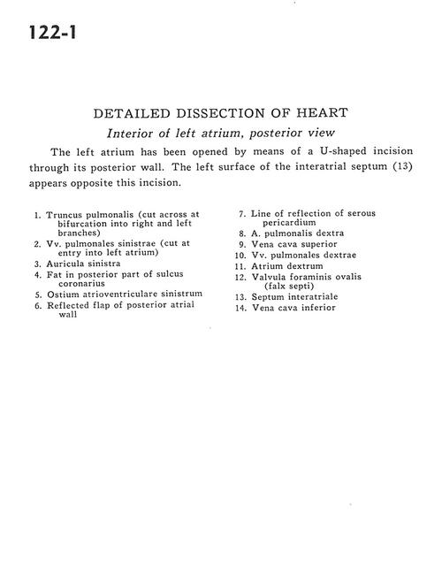
Detailed dissection of heart
Interior of left atrium, posterior view
The left atrium has been opened by means of a U-shaped incision through its posterior wall. The left surface of the interatrial septum (13) appears opposite this incision.
- Pulmonary trunk (cut across at bifurcation into right and left branches)
- Left pulmonary veins (cut at entry into left atrium)
- Left auricle
- Fat in posterior part of coronary sulcus
- Left atrioventricular opening
- Reflected flap of posterior atrial wall
- Line of reflection of serous pericardium
- Right pulmonary artery
- Superior vena cava
- Right pulmonary veins
- Right atrium
- Valve of foramen ovale (falx septi)
- Interatrial septum
- Inferior vena cava