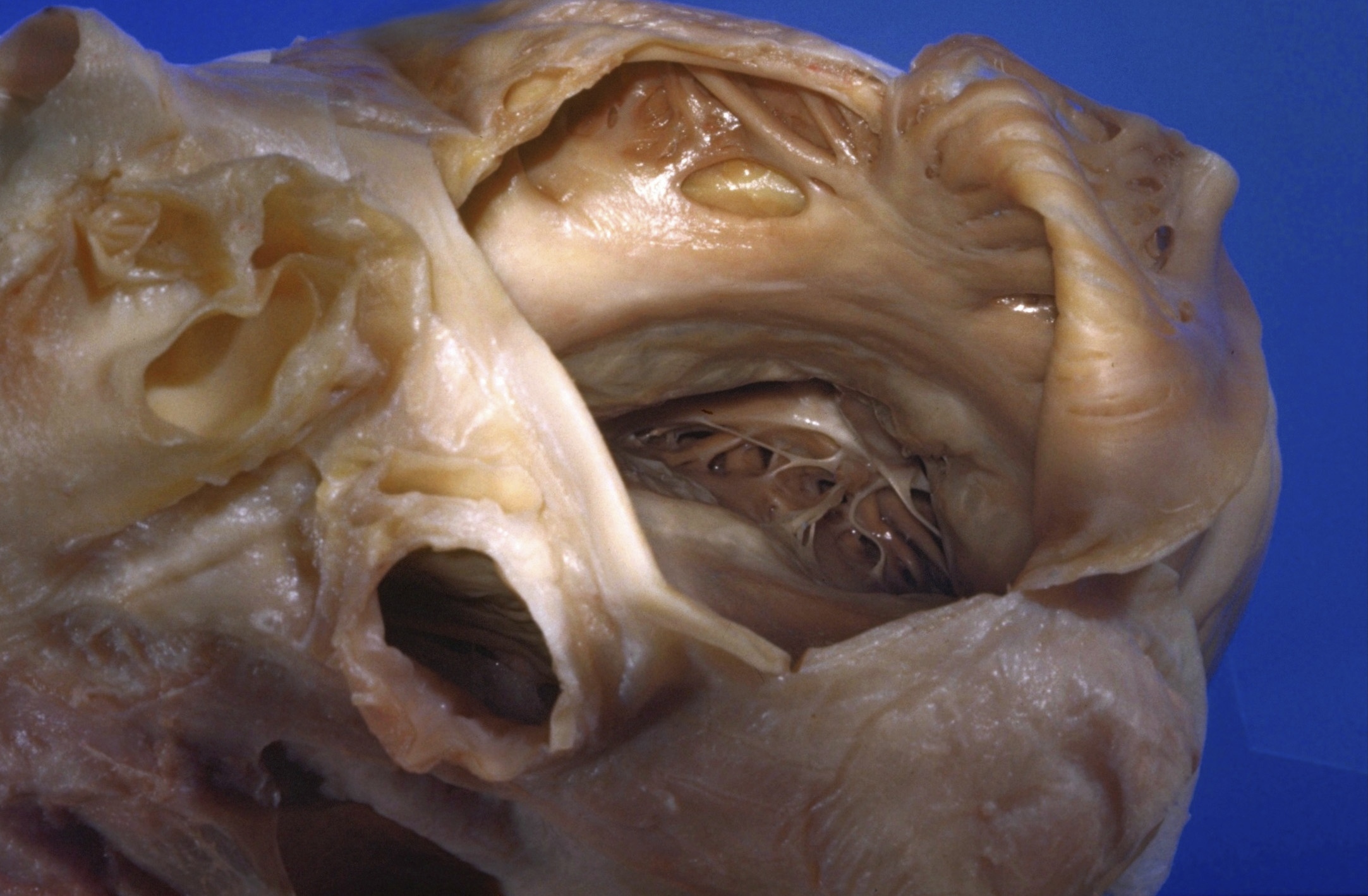
Detailed dissection of heart
Tricuspid valve viewed through right atrium
Stanford holds the copyright to the David L. Bassett anatomical images and has assigned
Creative Commons license Attribution-Share
Alike 4.0 International to all of the images.
For additional information regarding use and permissions,
please contact the Medical History Center.
Image #121-3
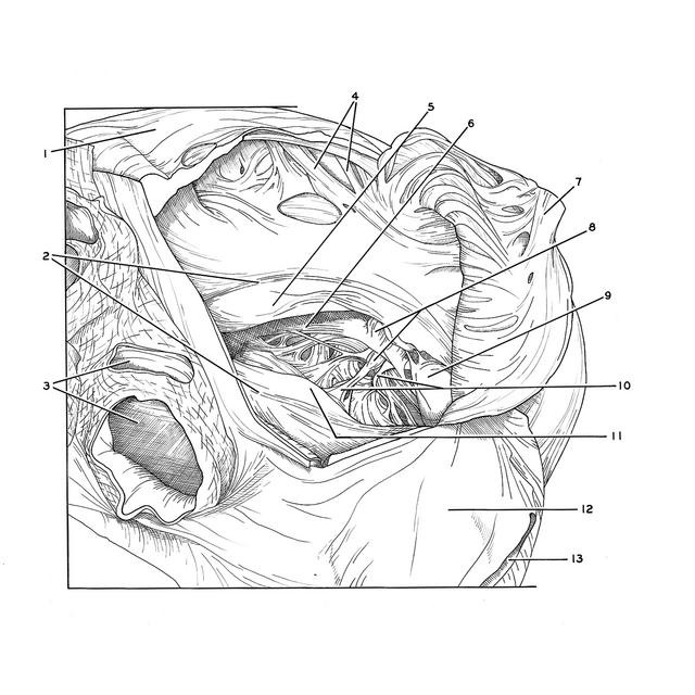
Detailed dissection of heart
Tricuspid valve viewed through right atrium
The view is directed into the cavity of the right ventricle through the opened right atrium and the ostium (2) of the tricuspid valve.
- Right auricle
- Margins of right atrioventricular opening
- Right pulmonary veins (cut across)
- Pectinate muscles
- Anterior cusp tricuspid valve
- Septomarginal trabecula (moderator band)
- Crista terminalis (in reflected part of atrial wall)
- Papillary muscles
- Posterior cusp tricuspid valve
- Chordae tendineae
- Septal (medial) cusp of tricuspid valve
- External surface of right atrium
- Inferior vena cava (cut off)