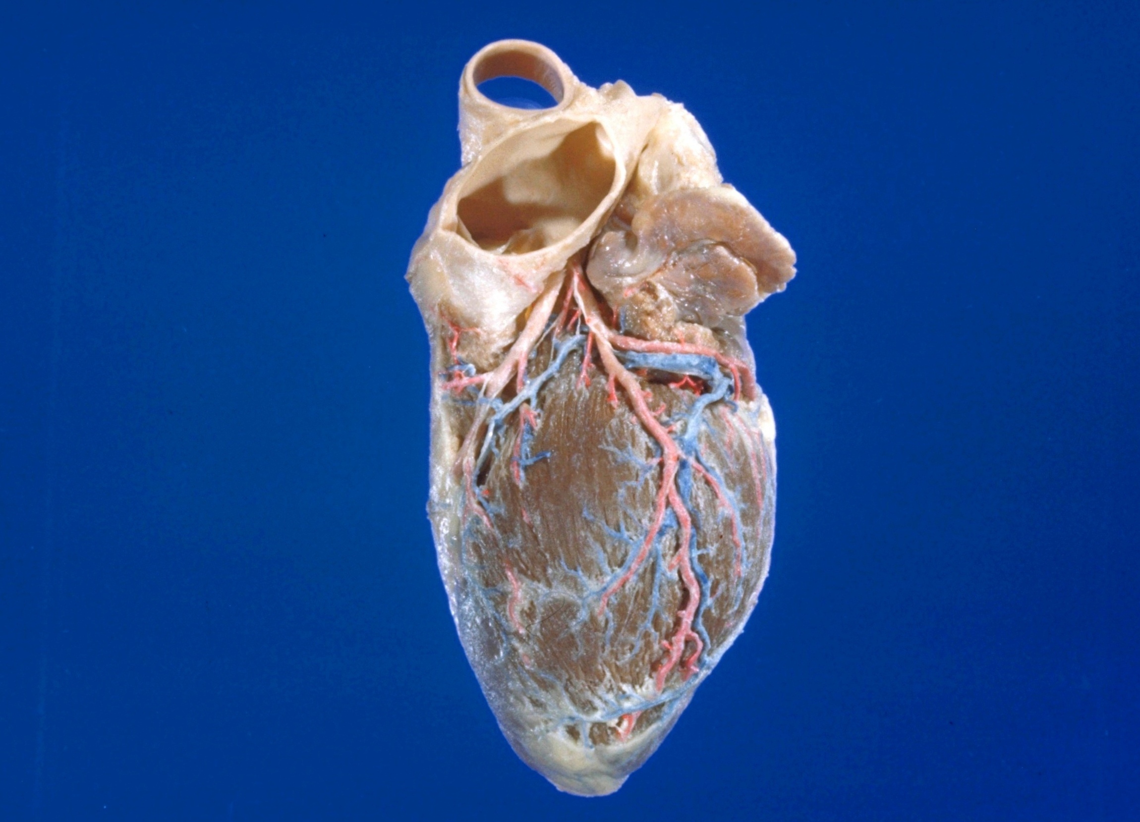
Detailed dissection of heart
Coronary vessels viewed from left side
Stanford holds the copyright to the David L. Bassett anatomical images and has assigned
Creative Commons license Attribution-Share
Alike 4.0 International to all of the images.
For additional information regarding use and permissions,
please contact the Medical History Center.
Image #119-4
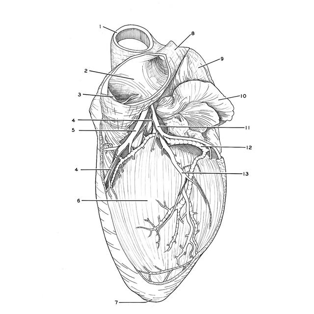
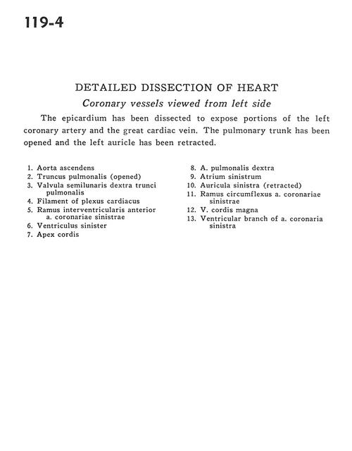
Detailed dissection of heart
Coronary vessels viewed from left side
The epicardium has been dissected to expose portions of the left coronary artery and the great cardiac vein. The pulmonary trunk has been opened and the left auricle has been retracted.
- Ascending aorta
- Pulmonary trunk (opened)
- Right semilunar cusp pulmonary trunk
- Filament of cardiac plexus
- Anterior interventricular branch of left coronary artery
- Left ventricle
- Apex of heart
- Right pulmonary artery
- Left atrium
- Left auricle (retracted)
- Circumflex branch of left coronary artery
- Great cardiac vein
- Ventricular branch of left coronary artery