Dissection of pericardium and heart in situ
Lines of reflection of serous pericardium onto heart and great vessels
Stanford holds the copyright to the David L. Bassett anatomical images and has assigned
Creative Commons license Attribution-Share
Alike 4.0 International to all of the images.
For additional information regarding use and permissions,
please contact the Medical History Center.
Image #118-7
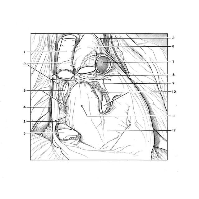
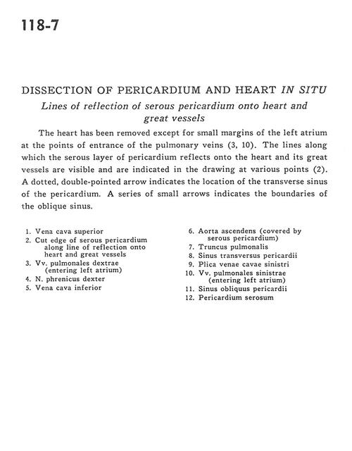
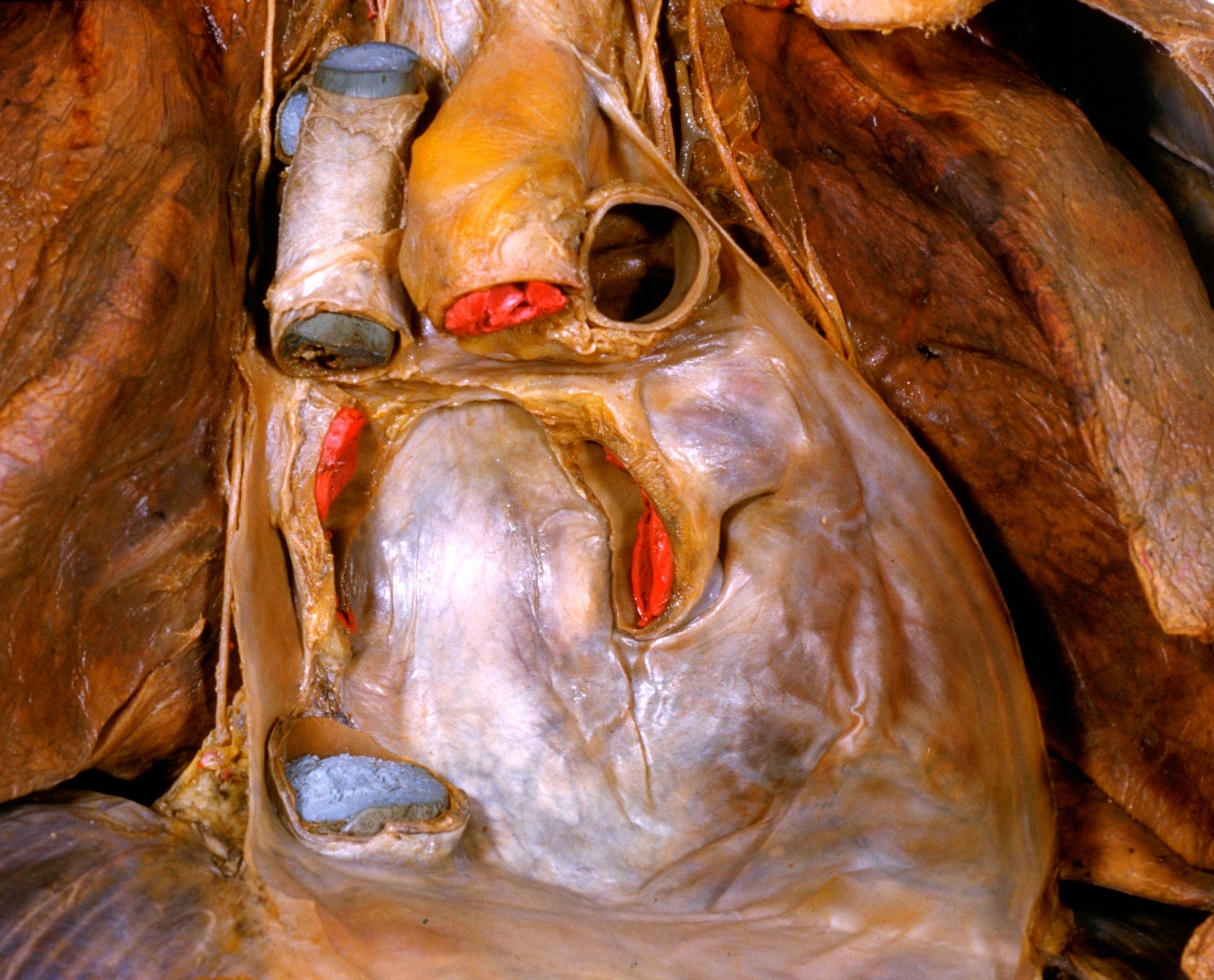
Dissection of pericardium and heart in situ
Lines of reflection of serous pericardium onto heart and great vessels
The heart has been removed except for small margins of the left atrium at the points of entrance of the pulmonary veins (3, 10). The lines along which the serous layer of pericardium reflects onto the heart and its great vessels are visible and are indicated in the drawing at various points (2). A dotted, double-pointed arrow indicates the location of the transverse sinus of the pericardium. A series of small arrows indicates the boundaries of the oblique sinus.
- Superior vena cava
- Cut edge of serous pericardium along line of reflection onto heart and great vessels
- Right pulmonary veins (entering left atrium)
- Right phrenic nerve
- Inferior vena cava
- Ascending aorta (covered by serous pericardium)
- Pulmonary trunk
- Transverse pericardial sinus
- Left fold of vena cava
- Left pulmonary veins (entering left atrium)
- Oblique pericardial sinus
- Serous pericardium