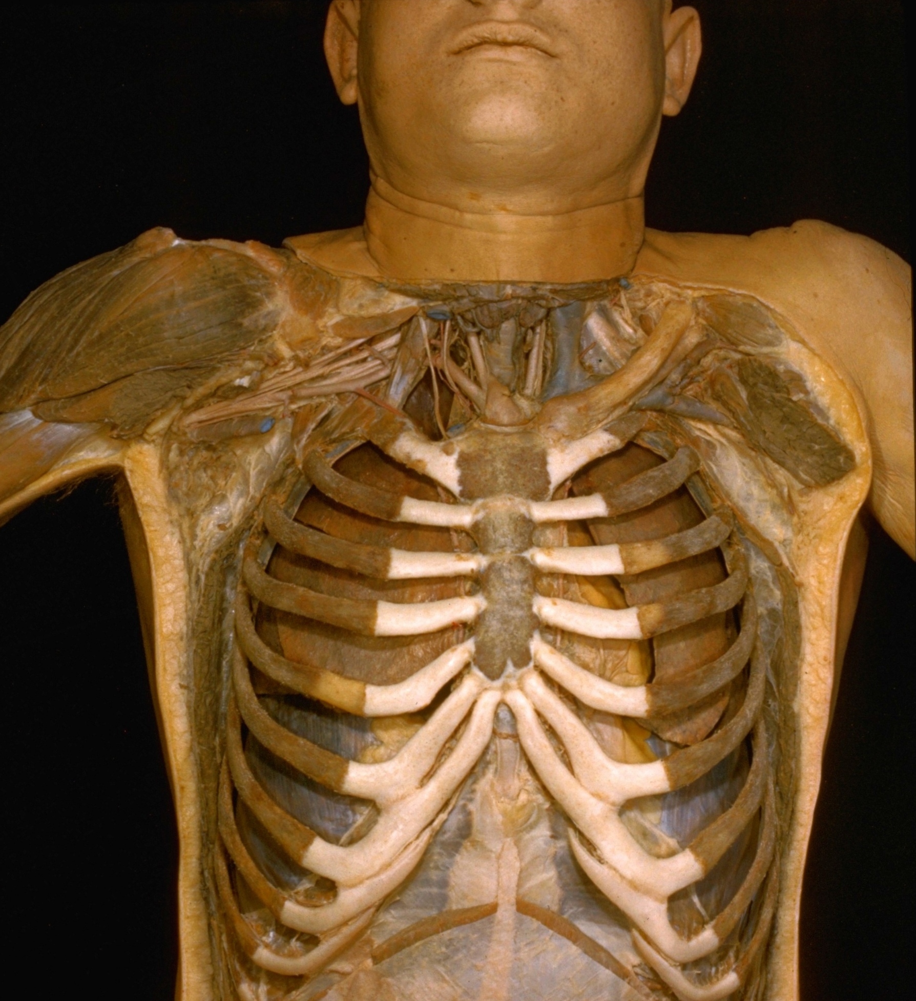
Thoracic viscera in situ
Anterior view of thoracic contents, rib cage intact
Stanford holds the copyright to the David L. Bassett anatomical images and has assigned
Creative Commons license Attribution-Share
Alike 4.0 International to all of the images.
For additional information regarding use and permissions,
please contact the Medical History Center.
Image #116-2

Thoracic viscera in situ
Anterior view of thoracic contents, rib cage intact
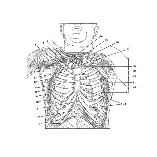
- Upper pointer: Phrenic nerve Lower pointer: Anterior scalene muscle
- Upper lobe right lung (in cupula pleurae, which has been opened)
- Subclavian artery
- Internal thoracic artery
- Upper lobe right lung
- Horizontal fissure right lung
- Middle lobe right lung
- Oblique fissure
- Inferior lobe right lung
- Diaphragmatic pleura
- Xiphoid process
- Sheath rectus abdominis muscle (posterior surface)
- Upper pointer: Rectus abdominis muscle (cut off) Lower pointer: Sheath of rectus abdominis muscle (anterior surface)
- Vagus nerve
- Trachea
- Left internal jugular vein
- Thymus
- Sternoclavicular joint
- Manubrium of sternum
- Upper lobe left lung
- Anterior mediastinum
- Oblique fissure
- Lower lobe left lung
- Cut edge of costal pleura at line of reflection onto mediastinum (upper pointer) and diaphragm (lower pointer)