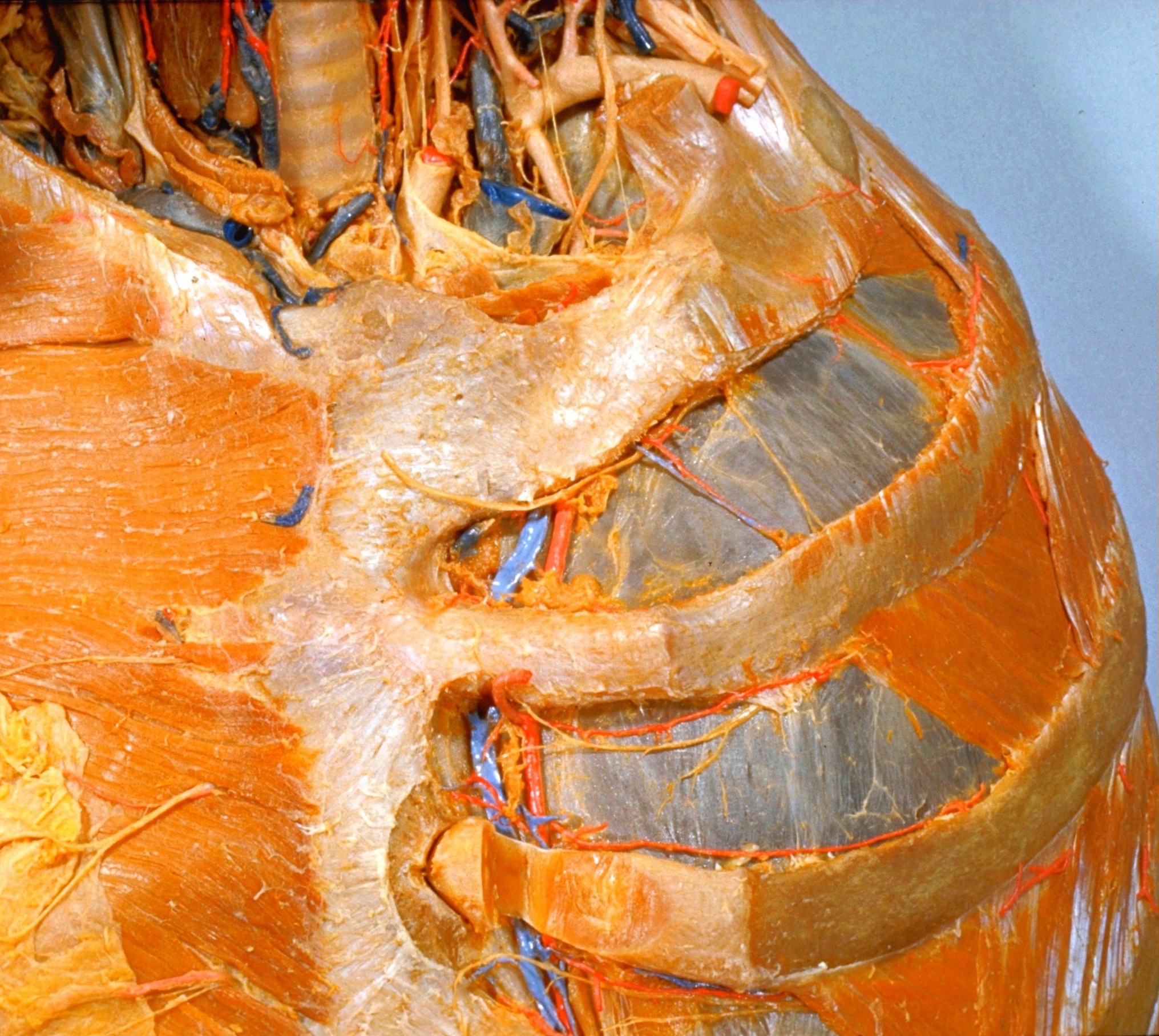
Dissection of breast and anterolateral thoracic wall
Vessels and nerves of first and second left intercostal spaces; Internal thoracic vessels; sternal lymph nodes; sternocostal joint
Stanford holds the copyright to the David L. Bassett anatomical images and has assigned
Creative Commons license Attribution-Share
Alike 4.0 International to all of the images.
For additional information regarding use and permissions,
please contact the Medical History Center.
Image #115-6
Dissection of breast and anterolateral thoracic wall
Vessels and nerves of first and second left intercostal spaces; Internal thoracic vessels; sternal lymph nodes; sternocostal joint
The costal part of the parietal pleura has been exposed in the anterior portions of the first and second intercostal spaces.
- Sternocleidomastoid muscle (cut off)
- Manubrium of sternum
- Pectoralis major muscle
- Anterior cutaneous branch intercostal nerve I
- Sternocostal ligament (pointer crosses sternal angle)
- Perforating branch internal thoracic artery
- Sternocostal joint III
- Sternocostal ligament (a delicate band in this specimen)
- Thyroid gland (right lobe)
- Trachea
- Left pointer: Thymus Right pointer: Common carotid artery
- Carotid sheath
- Left subclavian artery
- Anterior scalene muscle (cut off)
- Upper pointer: Internal thoracic artery (pointer near origin) Lower pointer: Costoclavicular ligament (cut off)
- Position of sternocostal joint I
- Costal pleura
- External intercostal muscle
- Sternal lymph nodes
- Internal intercostal muscle
- Intercostal nerve II
- Internal thoracic artery and vein
- Rib III
- Anterior intercostal branch internal thoracic artery
- Upper pointer: Costochondral junction Lower pointer: Costal cartilage III (note that a portion of the cartilage has been removed to allow the sternocostal joint to be opened)