Dorsal aspect of hand
Superficial branch of radial nerve at wrist, lateral view
Stanford holds the copyright to the David L. Bassett anatomical images and has assigned
Creative Commons license Attribution-Share
Alike 4.0 International to all of the images.
For additional information regarding use and permissions,
please contact the Medical History Center.
Image #109-6
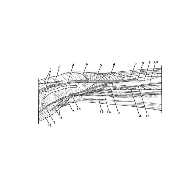
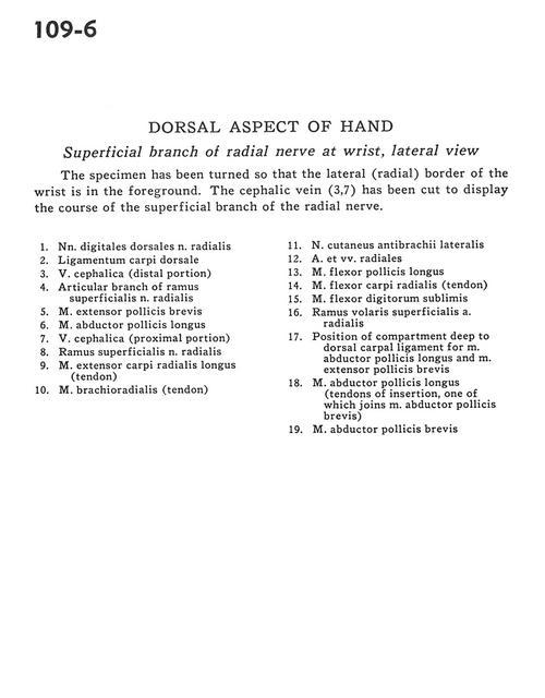
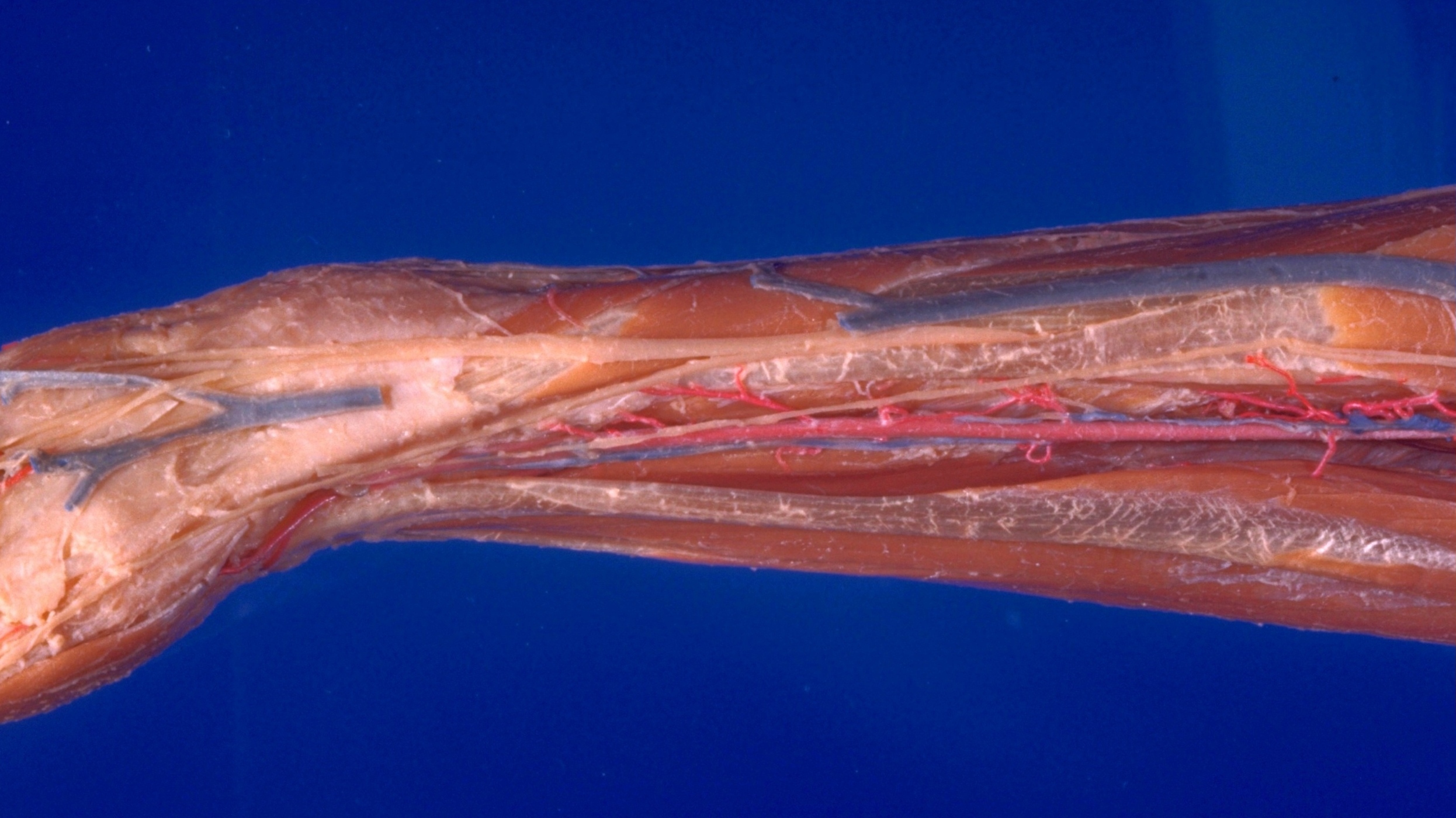
Dorsal aspect of hand
Superficial branch of radial nerve at wrist, lateral view
The specimen has been turned so that the lateral (radial) border of the wrist is in the foreground. The cephalic vein (3, 7) has been cut to display the course of the superficial branch of the radial nerve.
- Dorsal digital nerves of radial nerve
- Dorsal carpal ligament
- Cephalic vein (distal portion)
- Articular branch of superficial branch of radial nerve
- Extensor pollicis brevis muscle
- Abductor pollicis longus muscle
- Cephalic vein (proximal portion)
- Superficial branch of radial nerve
- Extensor carpi radialis longus muscle (tendon)
- Brachioradialis muscle (tendon)
- Lateral antebrachial cutaneous nerve
- Radial artery and veins
- Flexor pollicis longus muscle
- Flexor carpi radialis muscle (tendon)
- Flexor digitorum superficialis
- Superficial anterior branch radial artery
- Position of compartment deep to dorsal carpal ligament for abductor pollicis longus muscle and extensor pollicis brevis muscle
- Abductor pollicis longus muscle (tendons of insertion, one of which joins abductor pollicis brevis muscle)
- Abductor pollicis brevis muscle