Dorsal aspect of right forearm
Nerve supply to extensor carpi radialis brevis muscle
Stanford holds the copyright to the David L. Bassett anatomical images and has assigned
Creative Commons license Attribution-Share
Alike 4.0 International to all of the images.
For additional information regarding use and permissions,
please contact the Medical History Center.
Image #107-2
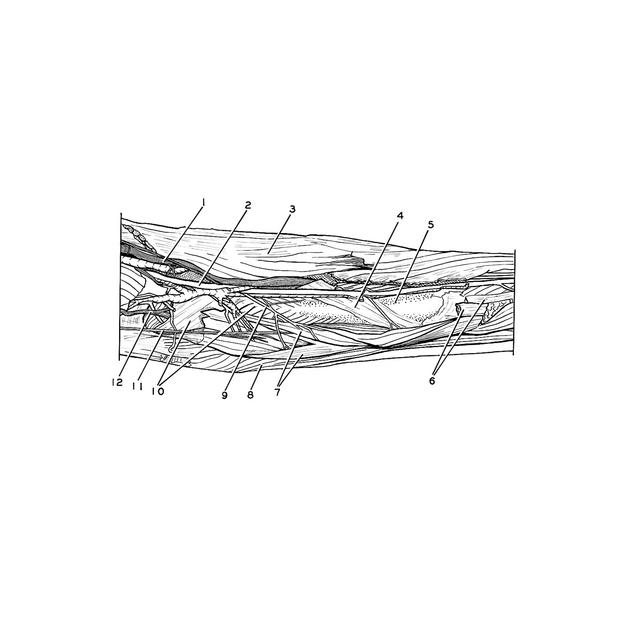
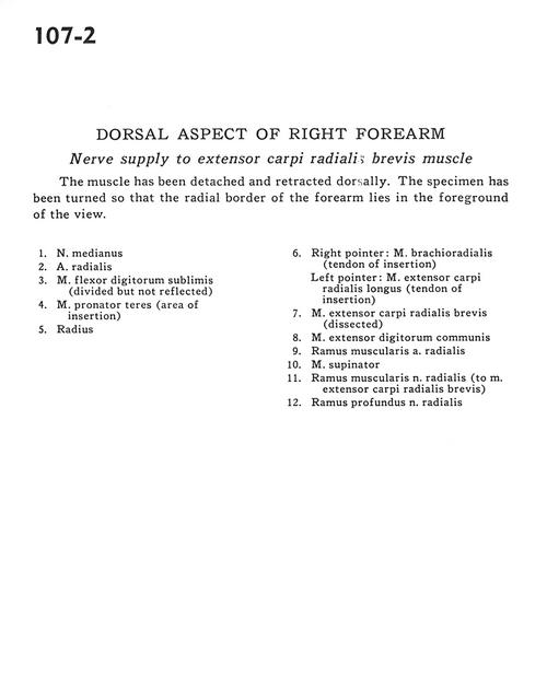
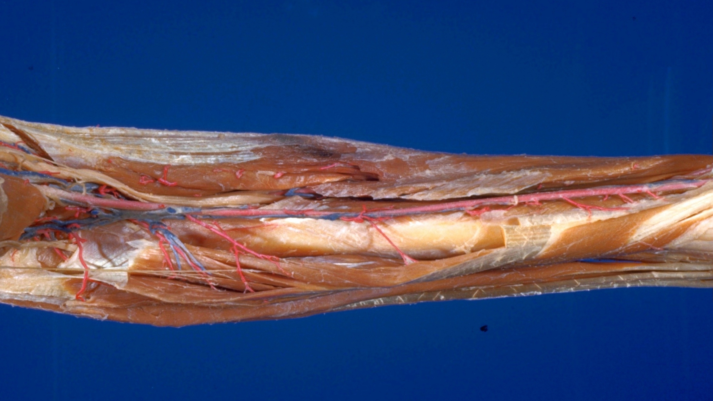
Dorsal aspect of right forearm
Nerve supply to extensor carpi radialis brevis muscle
The muscle has been detached and retracted dorsally. The specimen has been turned so that the radial border of the forearm lies in the foreground of the view.
- Median nerve
- Radial artery
- Flexor digitorum superficialis (divided but not reflected)
- Pronator teres muscle (area of insertion)
- Radius
- Right pointer: Brachioradialis muscle (tendon of insertion) Left pointer: Extensor carpi radialis longus muscle (tendon of insertion)
- Extensor carpi radialis brevis muscle (dissected)
- Common extensor digitorum muscle
- Muscular branch radial artery
- Supinator muscle
- Muscular branch of radial nerve (to extensor carpi radialis brevis muscle)
- Deep branch of radial nerve