Volar aspect of right hand
Relation of third and fourth lumbrical muscles to synovial sheaths of flexor tendons
Stanford holds the copyright to the David L. Bassett anatomical images and has assigned
Creative Commons license Attribution-Share
Alike 4.0 International to all of the images.
For additional information regarding use and permissions,
please contact the Medical History Center.
Image #102-1
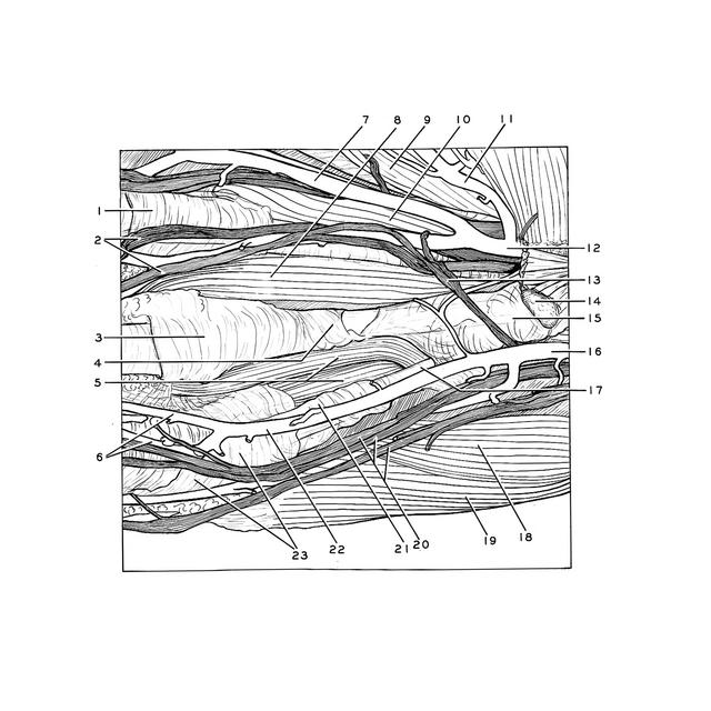
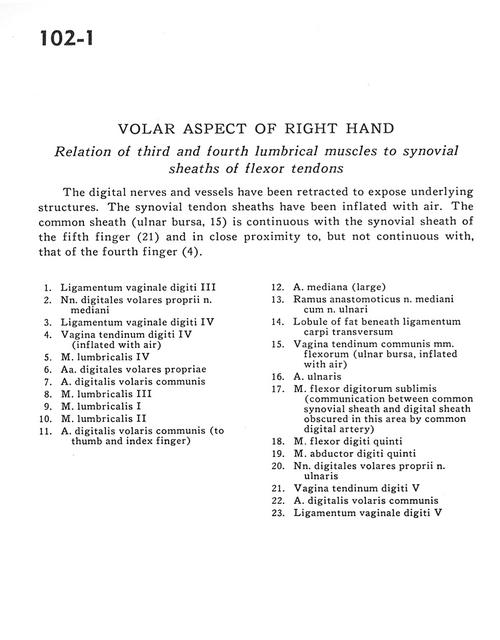
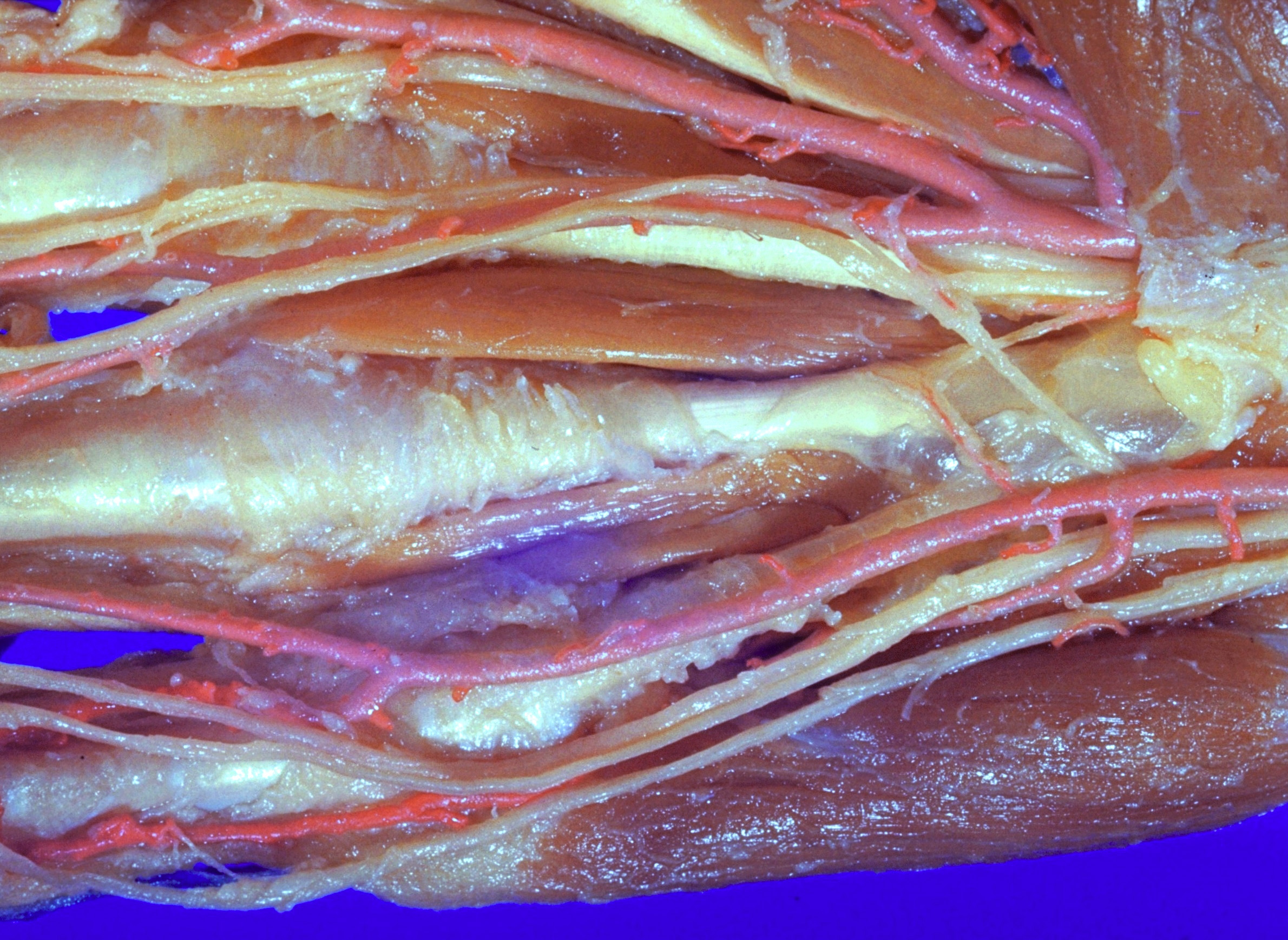
Volar aspect of right hand
Relation of third and fourth lumbrical muscles to synovial sheaths of flexor tendons
The digital nerves and vessels have been retracted to expose underlying structures. The synovial tendon sheaths have been inflated with air. The common sheath (ulnar bursa, 15) is continuous with the synovial sheath of the fifth finger (21) and in close proximity to, but not continuous with, that of the fourth finger (4).
- Ligament of digital sheath III
- Proper palmar digital nerves of median nerve
- Ligament of digital sheath IV
- Tendon of digital sheath IV (inflated with air)
- Lumbrical muscle IV
- Anterior proper digital arteries
- Anterior common digital artery
- Lumbrical muscle III
- Lumbrical muscle
- Lumbrical muscle II
- Anterior common digital artery (to thumb and index finger)
- Median artery (large)
- Anastomotic branch of median nerve with ulnar nerve
- Lobule of fat beneath transverse carpal ligament
- Sheath of common tendon of flexor muscles (ulnar bursa, inflated with air)
- Ulnar artery
- Flexor digitorum superficialis (communication between common synovial sheath and digital sheath obscured in this area by common digital artery)
- Flexor digiti minimi muscle
- Abductor digiti minimi muscle
- Proper palmar digital nerves of ulnar nerve
- Tendon of digital sheath V
- Anterior common digital artery
- Ligament of digital sheath V