Elbow joint
Articular cavity opened, posterior view
Stanford holds the copyright to the David L. Bassett anatomical images and has assigned
Creative Commons license Attribution-Share
Alike 4.0 International to all of the images.
For additional information regarding use and permissions,
please contact the Medical History Center.
Image #100-6
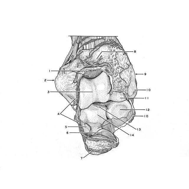
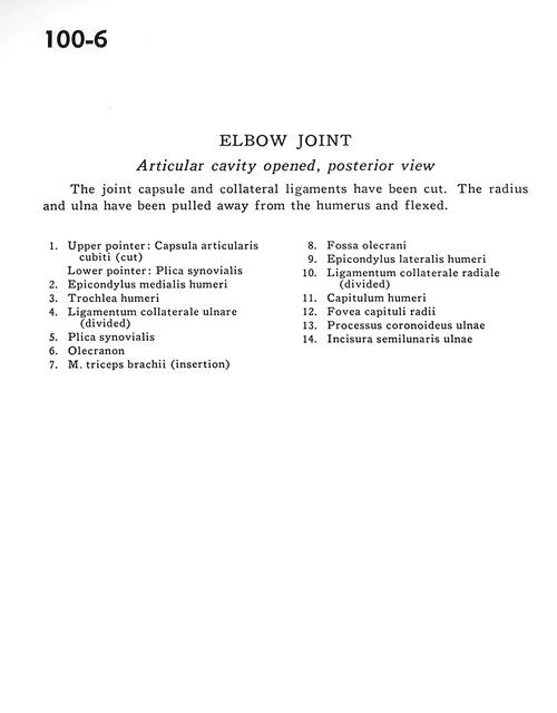
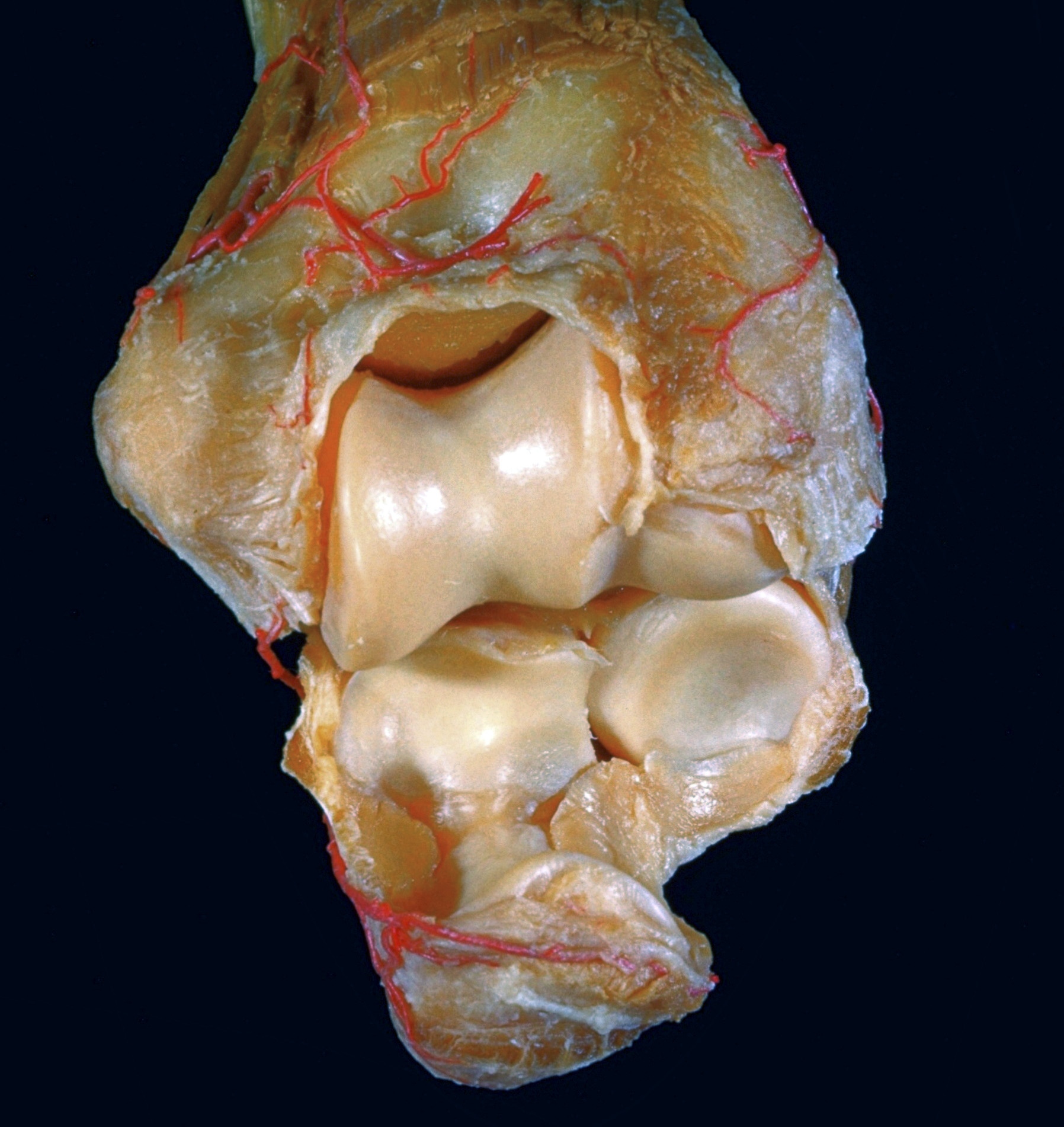
Elbow joint
Articular cavity opened, posterior view
The joint capsule and collateral ligaments have been cut. The radius and ulna have been pulled away from the humerus and flexed.
- Upper pointer: Cubital joint capsule (cut) Lower pointer: Synovial fold
- Medial epicondyle of humerus
- Trochlea of humerus
- Ulnar collateral ligament (divided)
- Synovial fold
- Olecranon
- Triceps brachii muscle (insertion)
- Olecranon fossa
- Lateral epicondyle of humerus
- Radial collateral ligament (divided)
- Head of humerus
- Fovea of head of radius
- Coronoid process of ulna
- Semilunar notch of ulna