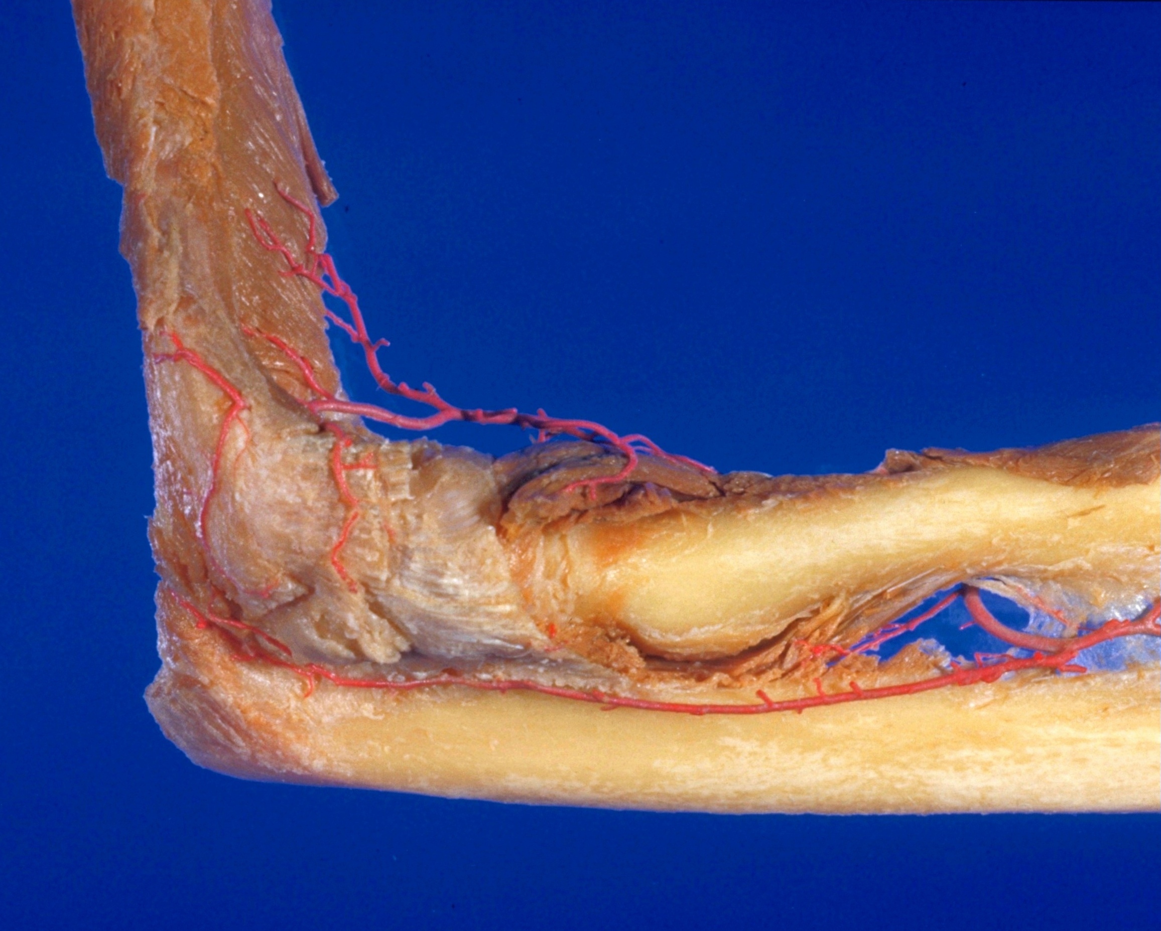
Elbow joint
Capsule and ligaments, lateral view of right elbow
Stanford holds the copyright to the David L. Bassett anatomical images and has assigned
Creative Commons license Attribution-Share
Alike 4.0 International to all of the images.
For additional information regarding use and permissions,
please contact the Medical History Center.
Image #100-4
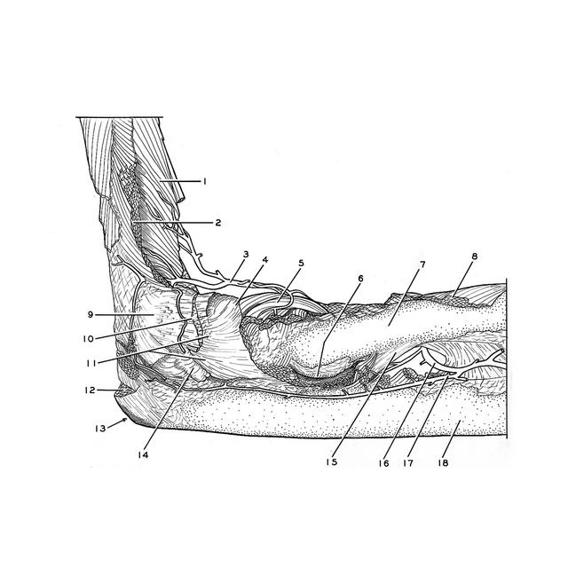
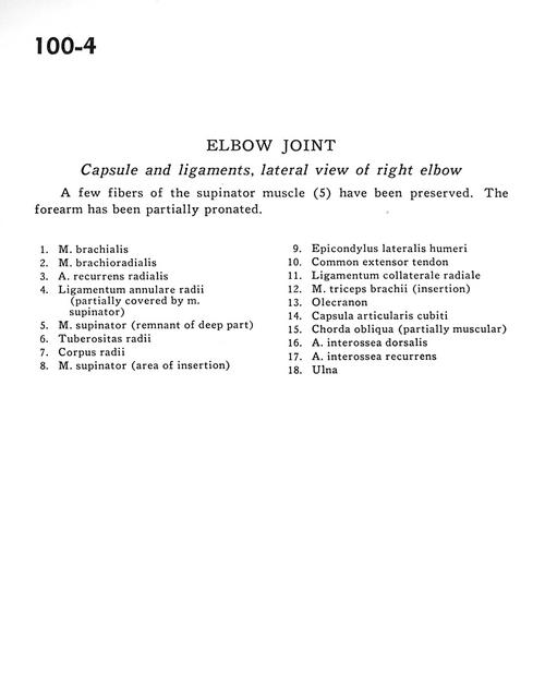
Elbow joint
Capsule and ligaments, lateral view of right elbow
A few fibres of the supinator muscle (5) have been preserved. The forearm has been partially pronated.
- Brachialis muscle
- Brachioradialis muscle
- Recurrent radial artery
- Annular ligament of radius (partially covered by supinator muscle)
- Supinator muscle (remnant of deep part)
- Tuberosity of radius
- Body of radius
- Supinator muscle (area of insertion)
- Lateral epicondyle of humerus
- Common extensor tendon
- Radial collateral ligament
- Triceps brachii muscle (insertion)
- Olecranon
- Cubital joint capsule
- Oblique cord (partially muscular)
- Dorsal interosseous artery
- Recurrent interosseous artery
- Ulna