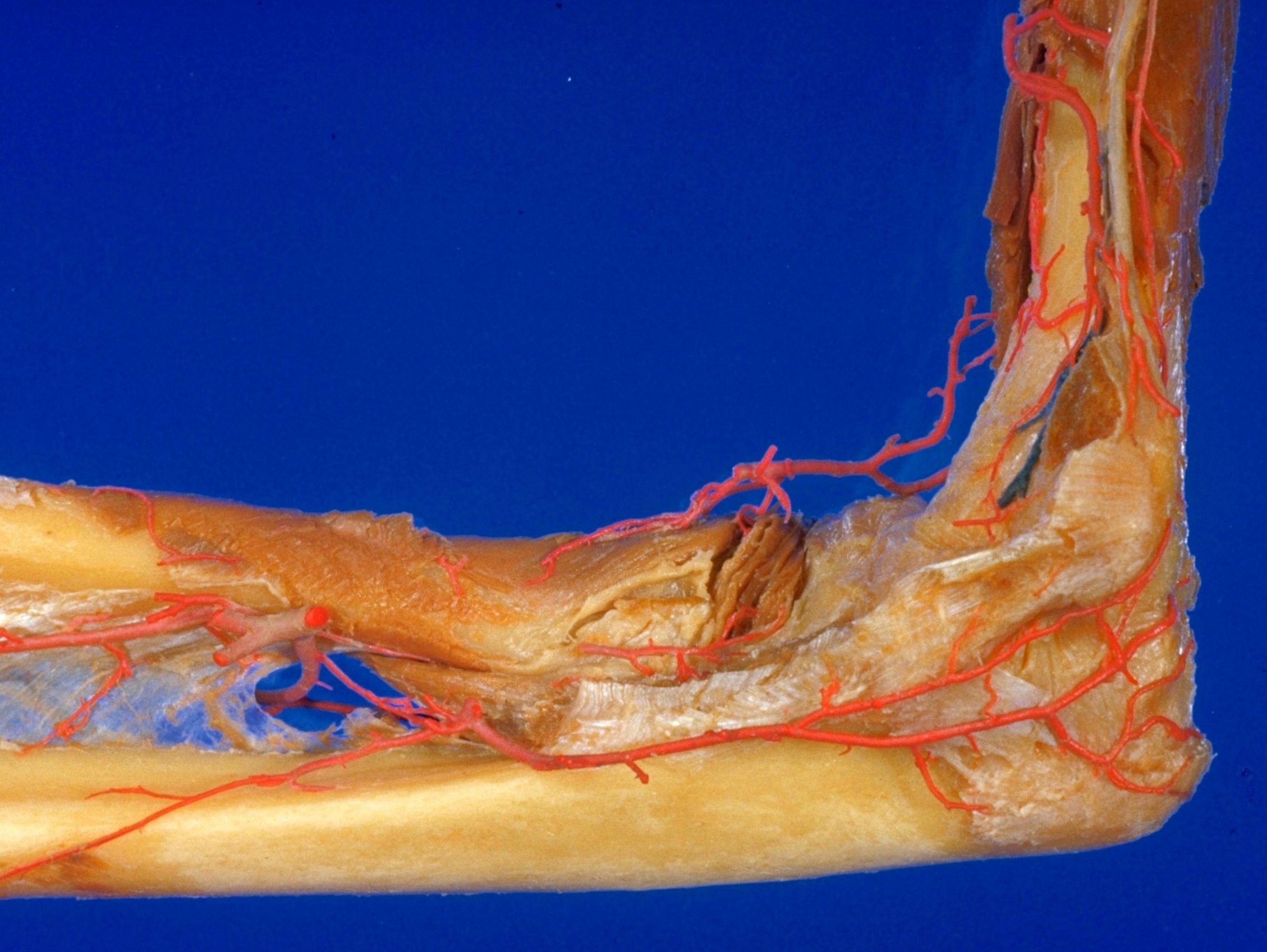
Elbow joint
Capsule and ligaments, medial view of right elbow
Stanford holds the copyright to the David L. Bassett anatomical images and has assigned
Creative Commons license Attribution-Share
Alike 4.0 International to all of the images.
For additional information regarding use and permissions,
please contact the Medical History Center.
Image #100-3
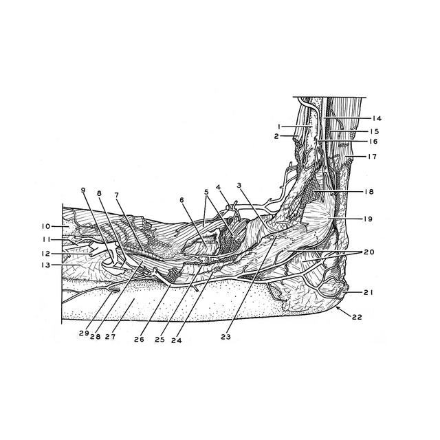
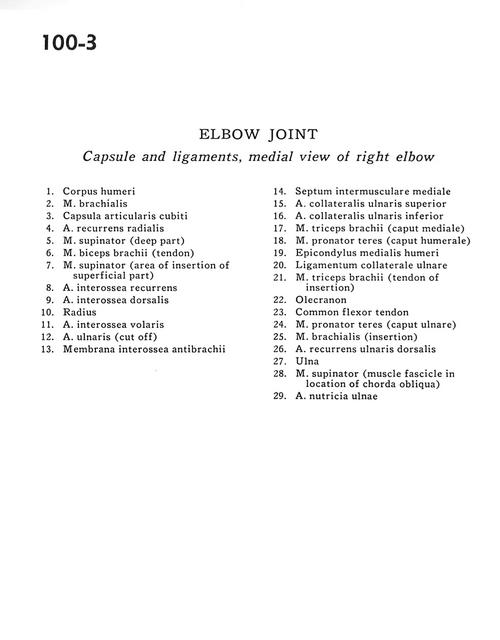
Elbow joint
Capsule and ligaments, medial view of right elbow
- Body of humerus
- Brachialis muscle
- Cubital joint capsule
- Recurrent radial artery
- Supinator muscle (deep part)
- Biceps brachii muscle (tendon)
- Supinator muscle (area of insertion of superficial part)
- Recurrent interosseous artery
- Dorsal interosseous artery
- Radius
- AnteriOr interosseous artery
- Ulnar artery (cut off)
- Antebrachial interosseous membrane
- Medial intermuscular septum:
- Superior ulnar collateral artery
- Inferior ulnar collateral artery
- Triceps brachii muscle (medial head)
- Pronator teres muscle (humeral head)
- Medial epicondyle of humerus
- Ulnar collateral ligament
- Triceps brachii muscle (tendon of insertion)
- Olecranon
- Common flexor tendon
- Pronator teres muscle (ulnar head)
- Brachialis muscle (insertion)
- Dorsal recurrent ulnar artery
- Ulna
- Supinator muscle (muscle fascicle in location of chorda obliqua)
- Nutritive artery of ulna