Exploration of the meninges and brain in situ
Lateral view of dura mater and meningeal vessels
Stanford holds the copyright to the David L. Bassett anatomical images and has assigned
Creative Commons license Attribution-Share
Alike 4.0 International to all of the images.
For additional information regarding use and permissions,
please contact the Medical History Center.
Image #1-1
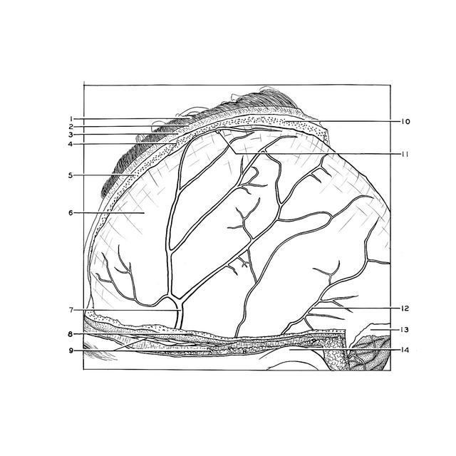
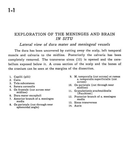
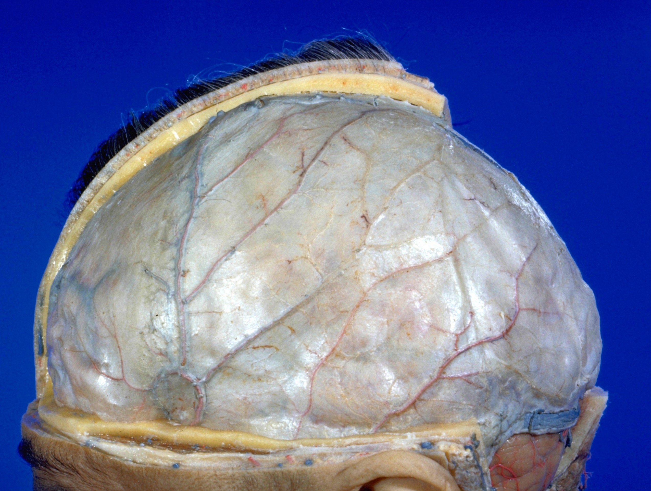
Exploration of the meninges and brain in situ
Lateral view of dura mater and meningeal vessels
The dura has been uncovered by cutting away the scalp, left temporal muscle and calvaria to the midline. Posteriorly the calvaria has been completely removed. The transverse sinus (13) is opened and the cerebellum exposed below it. A cross section of the scalp and the bones of the cranium can be seen at the margins of the dissection.
- Hair
- Skin
- Superficial fascia
- Coronal suture
- Frontal bone (cut across near midline)
- Dura mater
- Anterior branch of Middle meningeal artery
- Parietal bone (cut through near sphenoidal angle)
- Temporalis muscle (cut across) and Superficial temporal artery (cut across)
- Parietal bone (cut through near midline)
- Arachnoid granulations
- Posterior branch of middle meningeal artery
- Transverse sinus
- Auricle