X CLOSE

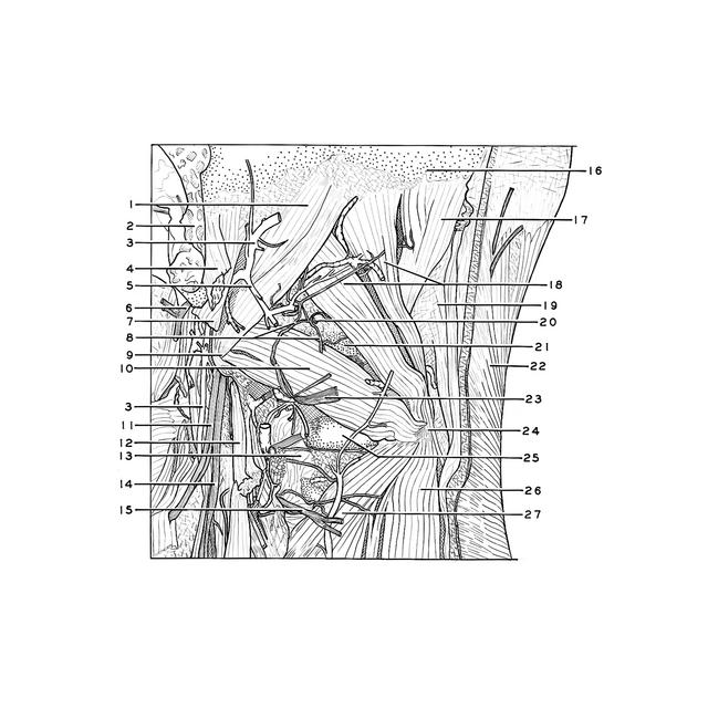
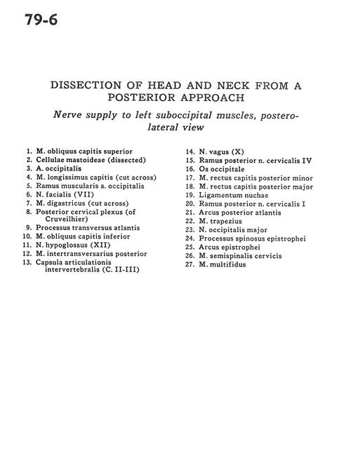
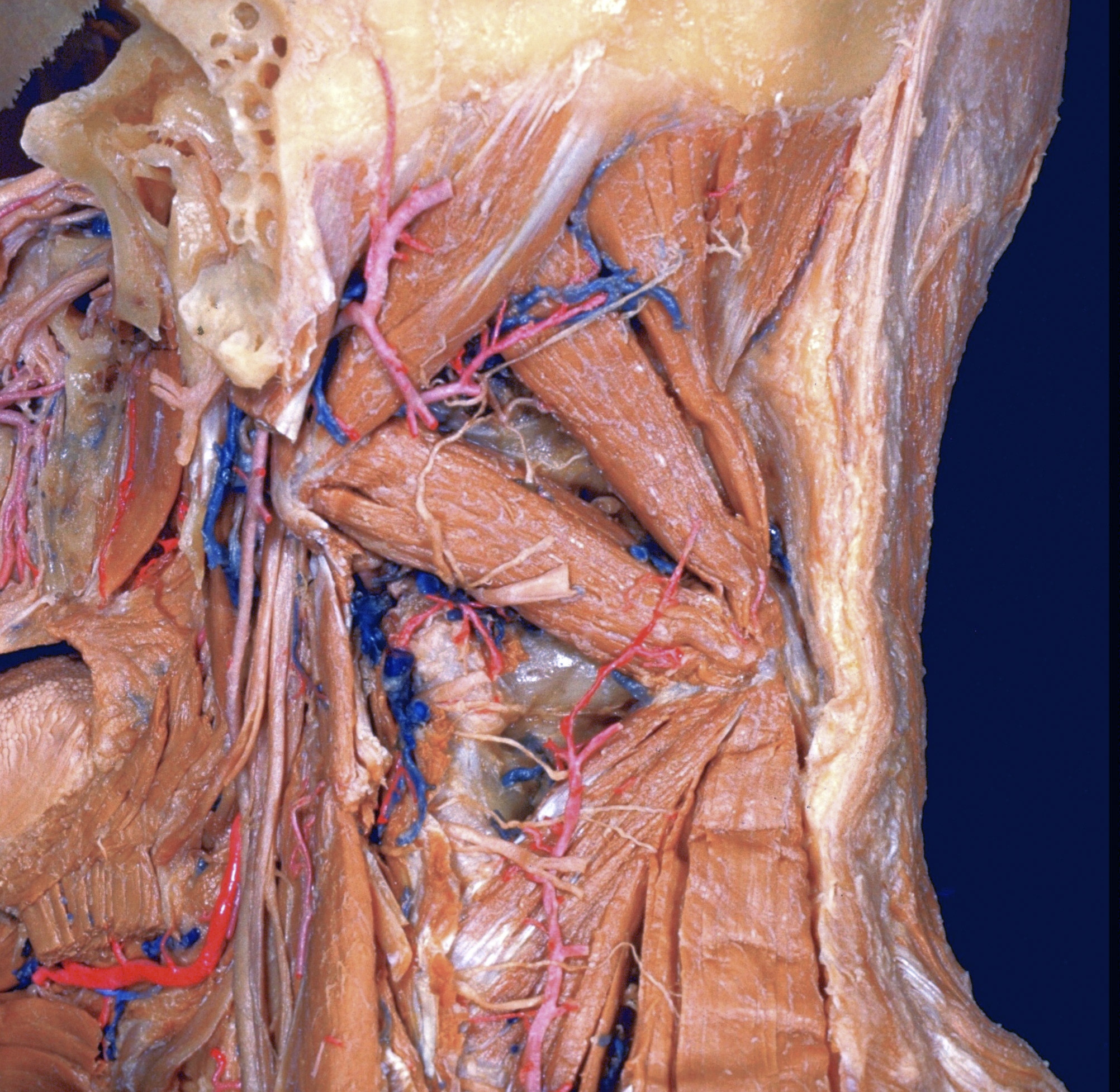
Dissection of head and neck from a posterior approach

Nerve supply to left suboccipital muscles, posterolateral view

Stanford holds the copyright to the David L. Bassett anatomical images and has assigned Creative Commons license Attribution-Share Alike 4.0 International to all of the images.
For additional information regarding use and permissions, please contact Dr. Drew Bourn at dbourn@stanford.edu.

Image #79-6
Bassett ImageDiagram Image
Legend Image
Dissection of head and neck from a posterior approach
Nerve supply to left suboccipital muscles, posterolateral view
1 . Superior oblique capitis muscle
2 . Mastoid cells (dissected)
3 . Occipital artery
4 . Longus capitis muscle (cut across)
5 . Muscular branch occipital artery
6 . Facial nerve (VII)
7 . Digastric muscle (cut across)
8 . Posterior cervical plexus (of Cruveilhier)
9 . Transverse process atlas
10 . Inferior oblique capitis muscle
11 . Hypoglossal nerve (XII)
12 . Intertransverse muscle (posterior)
13 . Intervertebral joint capsule (C. II-III)
14 . Vagus nerve (X)
15 . Posterior branch cervical nerve IV
16 . Occipital bone
17 . Minor posterior rectus capitis muscle
18 . Major posterior rectus capitis muscle
19 . Nuchal ligament
20 . Posterior branch cervical nerve I
21 . Posterior arch of atlas
22 . Trapezius muscle
23 . Greater occipital nerve
24 . Spinous process axis
25 . Arch of axis
26 . Semispinalis cervicis muscle
27 . Multifidus muscle