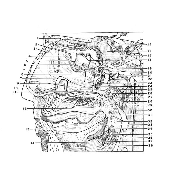
| 1
.
| Left pointer: Ethmoidal cell (opened) Right pointer: Olfactory bulb left |
| 2
.
| Superior nasal concha (partially resected) |
| 3
.
| Nasal septum (cut edge) |
| 4
.
| Ethmoidal infundibulum |
| 5
.
| Hiatus semilunaris |
| 6
.
| Ethmoidal bulla |
| 7
.
| Middle nasal concha (partially resected) |
| 8
.
| Middle nasal meatus |
| 9
.
| Upper pointer: Lacrimal fold Lower pointer: Ostium of nasolacrimal duct |
| 10
.
| Vestibule nasi and vibrissae (hair) |
| 11
.
| Inferior nasal concha (partially resected) |
| 12
.
| Upper pointer: Hard palate Lower pointer: Anterior palatine nerve |
| 13
.
| Gingiva |
| 14
.
| Mylohyoid muscle |
| 15
.
| Upper pointer: Optic nerve (II) left Lower pointer: Hypophysis |
| 16
.
| Sphenoid sinus left |
| 17
.
| Sphenoid sinus right (pointer on ostium) |
| 18
.
| Openings of ethmoidal cells (posterior) |
| 19
.
| Upper pointer: Posterior superior nasal branch of sphenopalatine ganglion Lower pointer: Posterior nasal septal artery |
| 20
.
| Upper pointer: Sphenopalatine foramen Lower pointer: Window cut in bone to expose pterygopalatine canal |
| 21
.
| Tunica mucosa pharynx |
| 22
.
| Pharyngeal branch of sphenopalatine ganglion |
| 23
.
| Upper pointer: Posterior lateral nasal artery Lower pointer: Posterior inferior (lateral) nasal branch of sphenopalatine ganglion |
| 24
.
| Greater palatine artery in the pterygopalatine canal |
| 25
.
| Upper pointer: Cartilaginous auditory tube Lower pointer: Mucosa |
| 26
.
| Palatine tensor veli muscle |
| 27
.
| Palatine levator veli muscle |
| 28
.
| Salpingopharyngeal muscle (note palatine branch of ascending palatine artery passing downward near pointer) |
| 29
.
| Superior constrictor pharyngis muscle |
| 30
.
| Prevertebral fascia |
| 31
.
| Uvula |
| 32
.
| Upper pointer: Glossopalatine muscle Lower pointer: Site of palatine tonsilla (resected) |
| 33
.
| Internal pterygoid muscle |
| 34
.
| Styloglossus muscle (cut across) |
| 35
.
| Submaxillary gland |
| 36
.
| Glossopharyngeal muscle (IX) |
| 37
.
| Hypoglossus nerve (XII) |
| 38
.
| Left pointer: Lingual nerve Right pointer: Submaxillary ganglion |
| *
.
| [Legend above restored translation from Latin] |