Dissection of nasal fossae, nasal pharynx, and palate
Relations of nasal passages, maxillary sinus, palate and sphenopalatine ganglion, left lateral view
Stanford holds the copyright to the David L. Bassett anatomical images and has assigned
Creative Commons license Attribution-Share Alike 4.0 International to all of the images.
For additional information regarding use and permissions,
please contact Dr. Drew Bourn at dbourn@stanford.edu.
Image #71-5
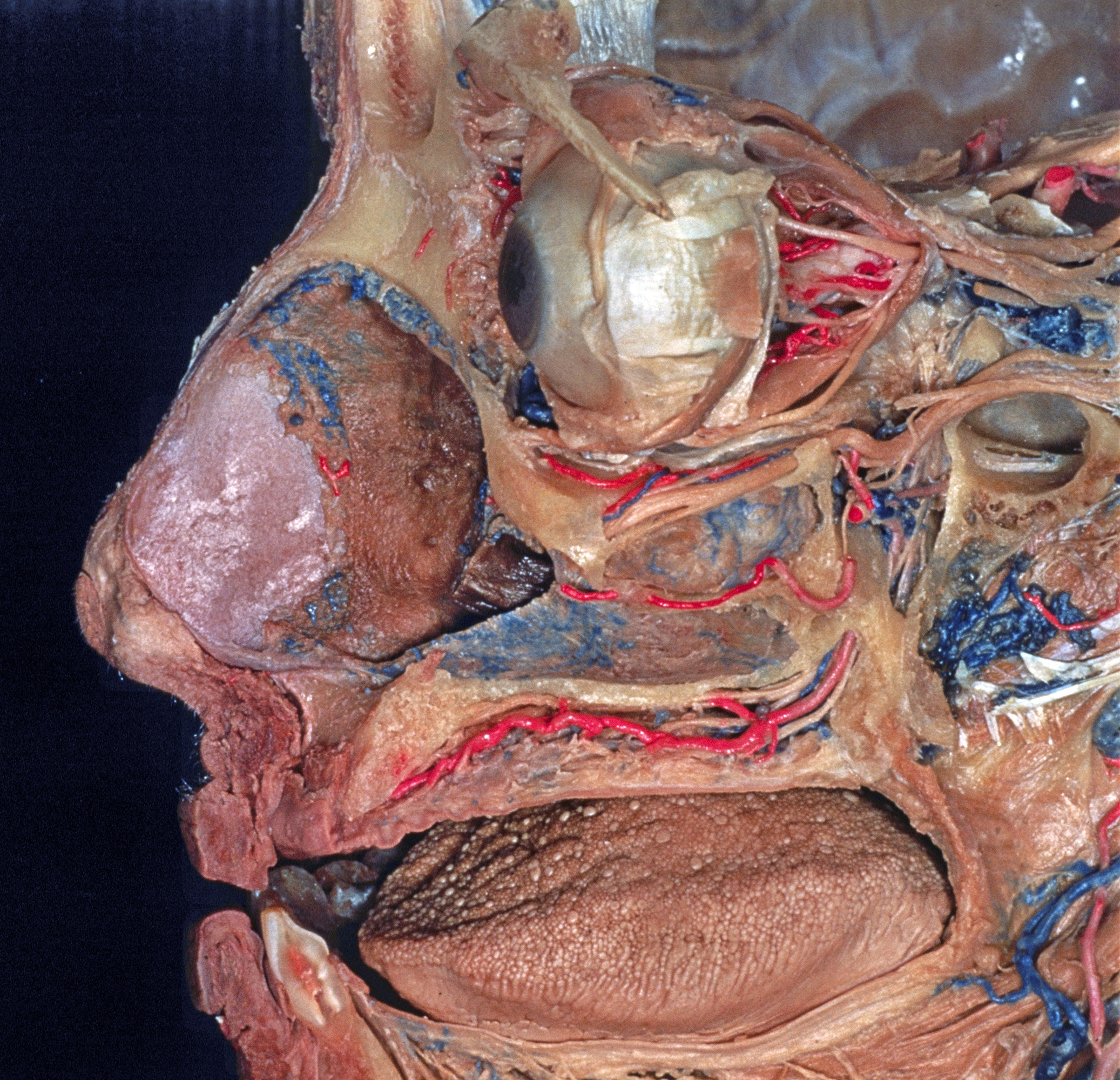 | 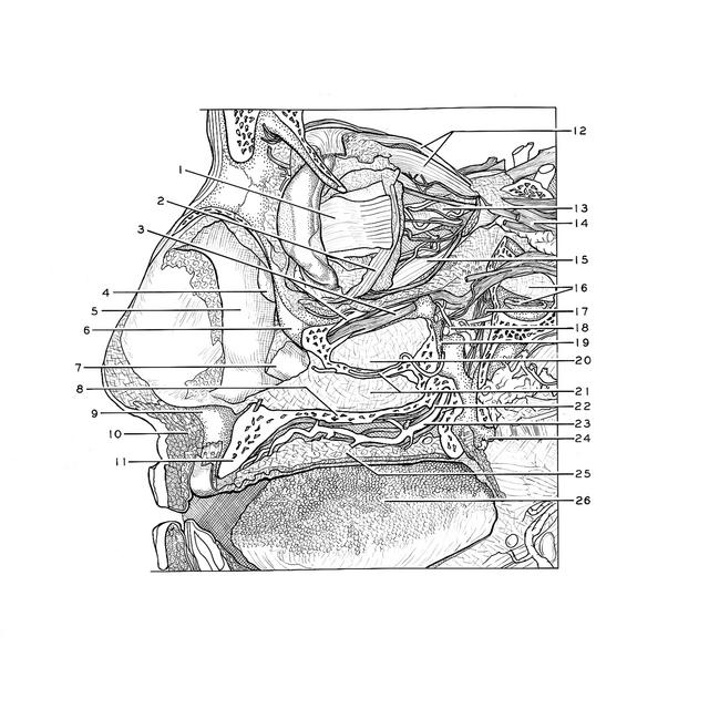 |
 | | Dissection of nasal fossae, nasal pharynx, and palate | | Relations of nasal passages, maxillary sinus, palate and sphenopalatine ganglion, left lateral view | | The zygomatic bone has been completely removed and the lateral bony walls of the maxillary sinus (20) and inferior nasal meatus (21) have been cut away. The sinus is unusually small. The connections of the sphenopalatine ganglion (17) have been dissected. | | 1
.
| Tendon of lateral rectus muscle | | 2
.
| Inferior oblique muscle | | 3
.
| Infraorbital nerve and artery within infraorbital canal (note branch of artery to inferior oblique muscle) | | 4
.
| Middle nasal concha | | 5
.
| Mucosa of nasal septum | | 6
.
| Infraorbital margin | | 7
.
| Inferior nasal concha (anterior tip resected) | | 8
.
| Hard palate | | 9
.
| Anterior nasal spine maxillae | | 10
.
| Muscles of upper lip (sectioned in midline) | | 11
.
| Alveolar process of maxilla (edentulous) | | 12
.
| Levator palpebrae superioris muscle and superior rectus muscle | | 13
.
| Sheath of optic nerve | | 14
.
| Ophthalmic nerve (V) | | 15
.
| Inferior rectus muscle | | 16
.
| Upper pointer: Sphenoid sinus (cut open) Lower pointer: Vidian nerve of pterygoid canal (bony canal opened) | | 17
.
| Sphenopalatine ganglion | | 18
.
| Terminal portion of internal maxillary artery (cut off) | | 19
.
| Superior posterior alveolar nerve | | 20
.
| Mucosal membrane maxillary sinus | | 21
.
| Superior posterior alveolar artery and mucosal membrane inferior nasal meatus | | 22
.
| Branches of anterior palatine nerve | | 23
.
| Greater palatine artery | | 24
.
| Remnant of lateral plate of pterygoid process | | 25
.
| Tunica palatine mucosa | | 26
.
| Tongue |
|
|


