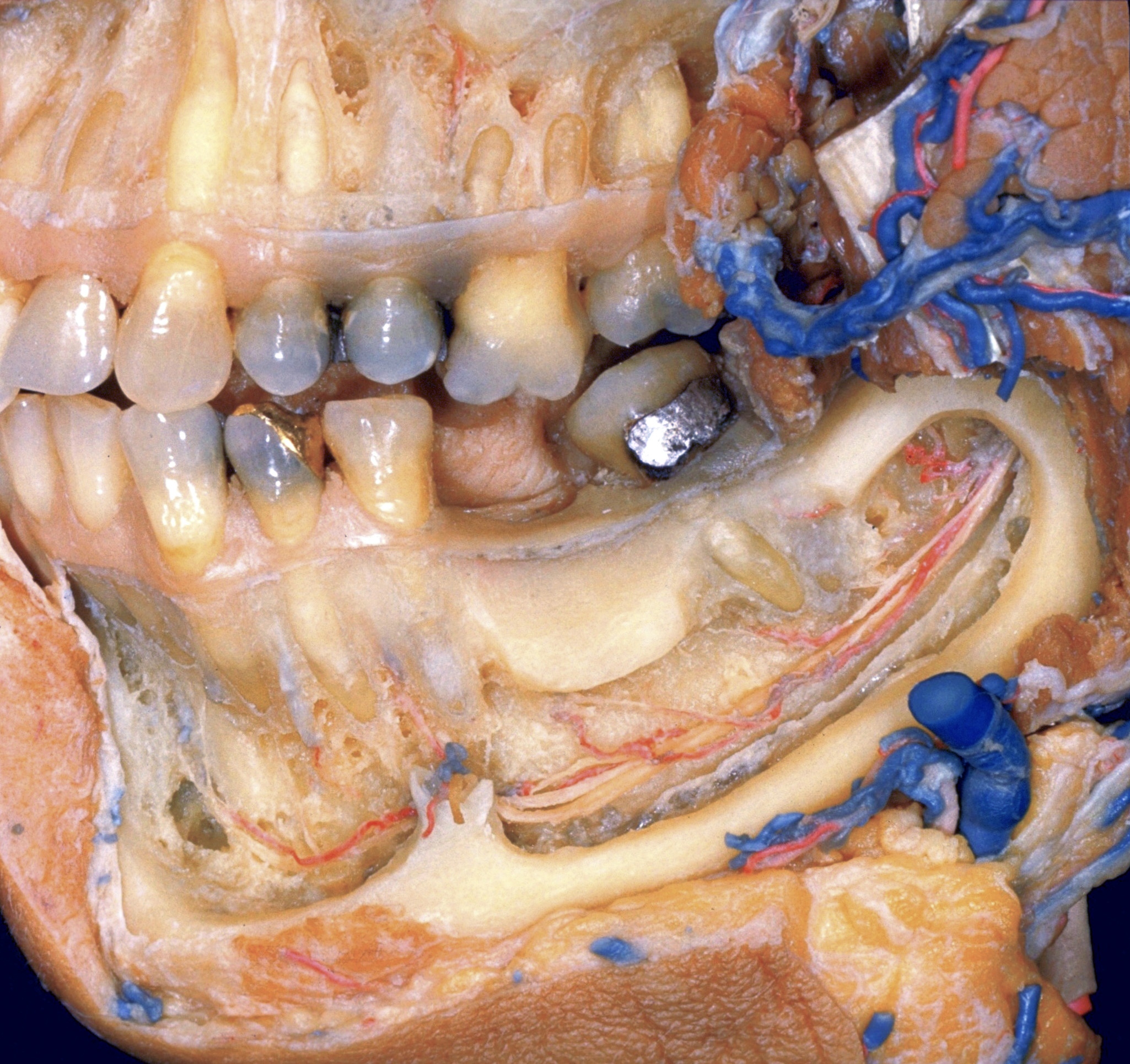
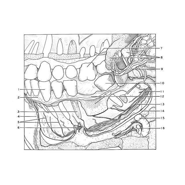
Nerves and blood vessels to lower teeth, left lateral view
For permissions information regarding the use of these images,
please contact Drew Bourn at dbourn@stanford.edu
Image #69-1
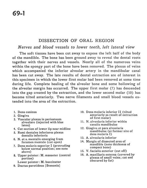
Dissection of oral region
Nerves and blood vessels to lower teeth, left lateral view
The soft tissues have been cut away to expose the left half of the body of the mandible. The bone has been ground away to reveal the dental roots together with their nerves and vessels. Nearly all of the numerous veins which accompanied the inferior alveolar artery in the mandibular canal has been cut away. The late results of dental extraction are of interest in this specimen in which the lower first molar had been removed at some time during life. Complete healing of the alveolar bone and some hollowing of the alveolar margin has occurred. The upper first-molar (7) has descended into the gap created by the extraction, and the lower second molar (10) has become tilted anteriorly. Two nerve filaments and small blood vessels extended into the area of the extraction.
1
.
Canine
2
.
Gingiva
3
.
Vascular plexus in alveolar periosteum (injected with blue latex)
4
.
Cut section of lower lip near midline
5
.
Inferior dental branches inferior dental plexus
6
.
Mental nerve and artery emerging from mental foramen (cut open)
7
.
Upper molar I (protruding below normal position see note above)