X CLOSE

Dissection of left orbit from a lateral approach

Eye sectioned vertically, optic nerve and papilla

Stanford holds the copyright to the David L. Bassett anatomical images and has assigned Creative Commons license Attribution-Share Alike 4.0 International to all of the images.
For additional information regarding use and permissions, please contact Dr. Drew Bourn at dbourn@stanford.edu.

Image #56-6
Bassett ImageDiagram Image
Legend Image
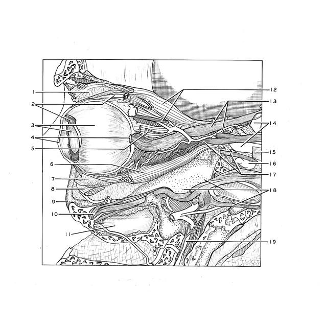
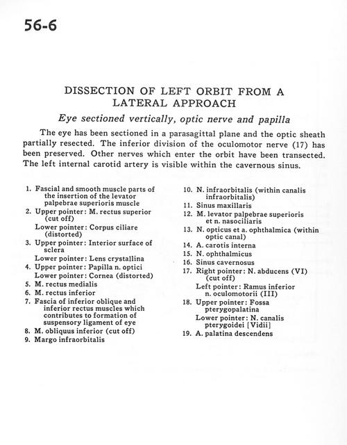
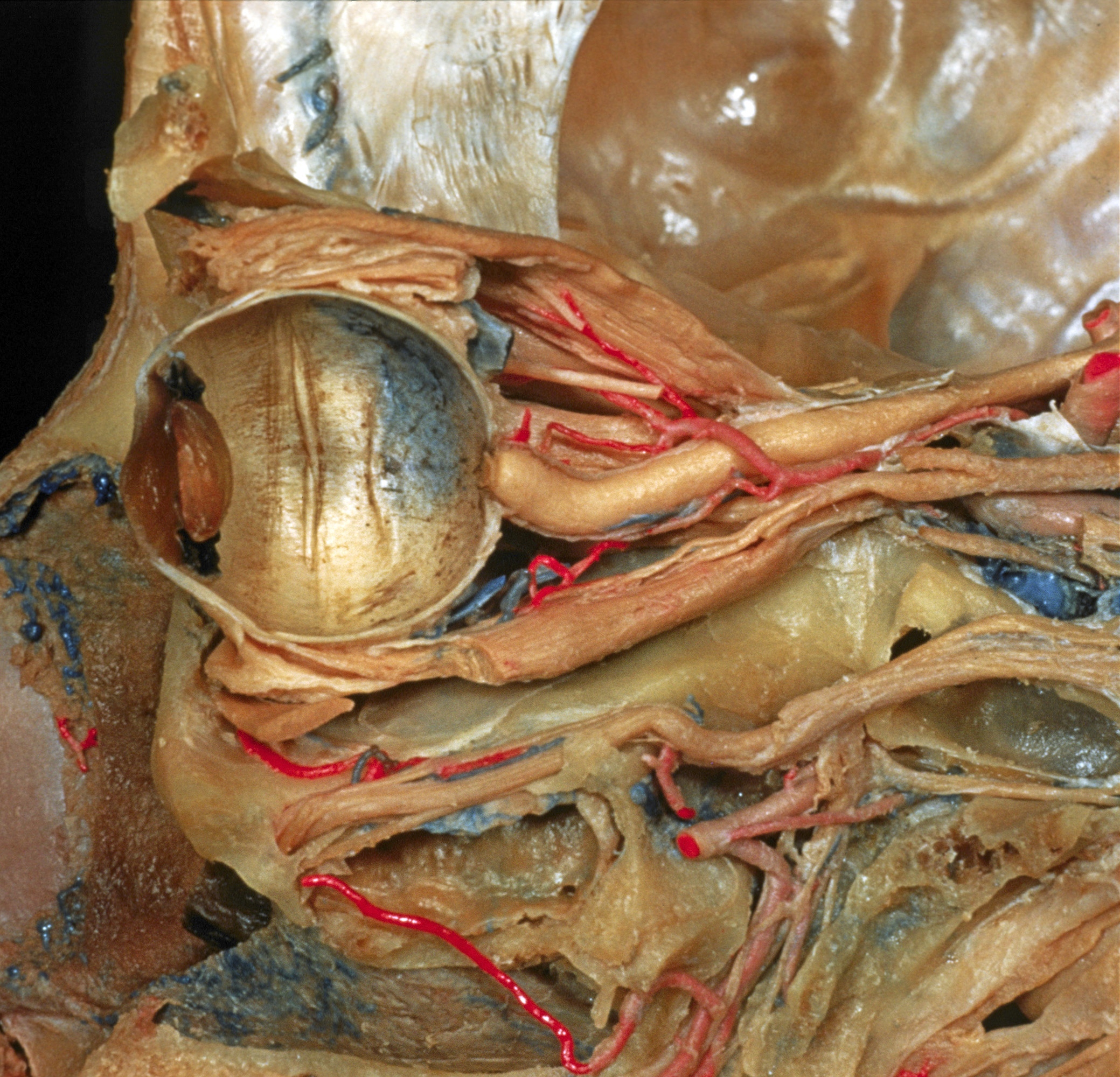
Dissection of left orbit from a lateral approach
Eye sectioned vertically, optic nerve and papilla
The eye has been sectioned in a parasagittal plane and the optic sheath partially resected. The inferior division of the oculomotor nerve(17) has been preserved. Other nerves which enter the orbit have been transected. The left internal carotid artery is visible withing the cavernous sinus.
1 . Fascial and smooth muscle parts of the insertion of the levator palpebrae superioris muscle
2 . Upper pointer: Superior rectus muscle (cut off) Lower pointer: Ciliary body (distorted)
3 . Upper pointer: Interior surface of sclera Lower pointer: Lens
4 . Upper pointer: Papilla optic nerve Lower pointer: Cornea (distorted)
5 . Medial rectus muscle
6 . Inferior rectus muscle
7 . Fascia of inferior oblique and inferior rectus muscles which contributes to formation of suspensory ligament of eye
8 . Inferior oblique muscle (cut off)
9 . Infraorbital margin
10 . Infraorbital nerve (within infraorbital canal)
11 . Maxillary sinus
12 . Levator palpebrae superioris muscle and nasociliary nerve
13 . Optic nerve and ophthalmic artery (within optic canal)
14 . Internal carotid artery
15 . Ophthalmic nerve
16 . Cavernous sinus
17 . Right pointer: Abducens nerve (VI) (cut off) Left pointer: Inferior branch oculomotor nerve (III)
18 . Upper pointer: Pterygopalatine fossa Lower pointer: Vidian nerve (of pterygoid canal)
19 . Descending palatine artery