X CLOSE

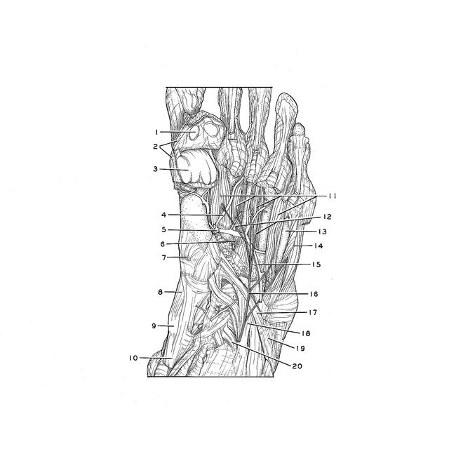
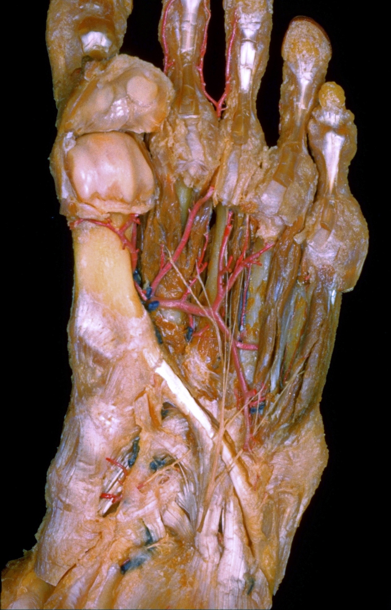
Dissection of plantar aspect of left foot

Nerve supply to dorsal interosseous muscles; interior of first metatarsophalangeal joint

Stanford holds the copyright to the David L. Bassett anatomical images and has assigned Creative Commons license Attribution-Share Alike 4.0 International to all of the images.
For additional information regarding use and permissions, please contact Dr. Drew Bourn at dbourn@stanford.edu.

Image #202-4
Bassett ImageDiagram Image
Legend Image
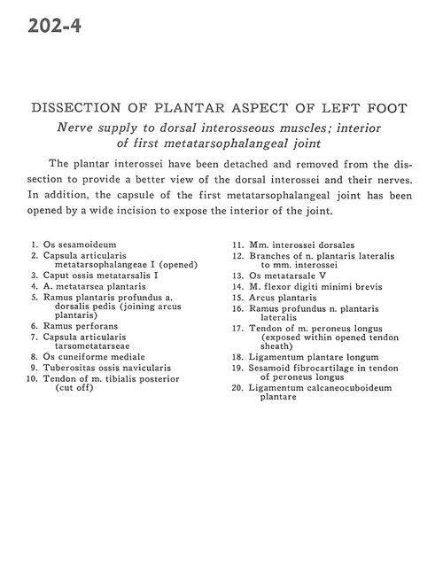
Dissection of plantar aspect of left foot
Nerve supply to dorsal interosseous muscles; interior of first metatarsophalangeal joint
The plantar interossei have been detached and removed from the dissection to provide a better view of the dorsal interossei and their nerves. In addition, the capsule of the first metatarsophalangeal joint has been opened by a wide incision to expose the interior of the joint.
1 . Sesamoid bone
2 . 1st metatarsophalangeal articular capsule (opened)
3 . Head of 1st metatarsal bone
4 . Plantar metatarsal artery
5 . Deep plantar branch of dorsalis pedis artery (joining plantar arch)
6 . Perforating branch
7 . Tarsometatarsal articular capsule
8 . Medial cuneiform bone
9 . Tuberosity of navicular bone
10 . Tendon of tibialis posterior muscle (cut off)
11 . Dorsal interosseous muscles
12 . Branches of lateral plantar nerve to interosseous muscle
13 . 5th metatarsal bone
14 . Flexor digiti minimi brevis muscle
15 . Plantar arch
16 . Deep branch lateral plantar nerve
17 . Tendon of peroneus longus muscle (exposed within opened tendon sheath)
18 . Long plantar ligament
19 . Sesamoid fibrocartilage in tendon of peroneus longus
20 . Calcaneocuboid plantar ligament