X CLOSE

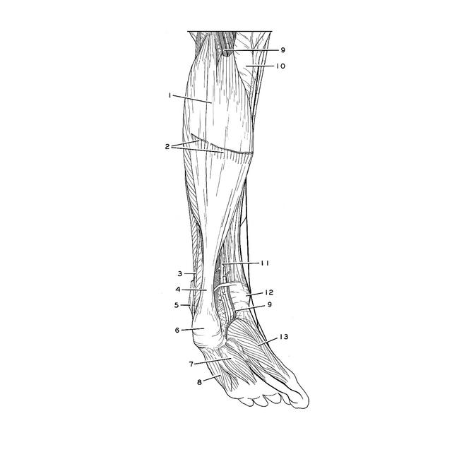
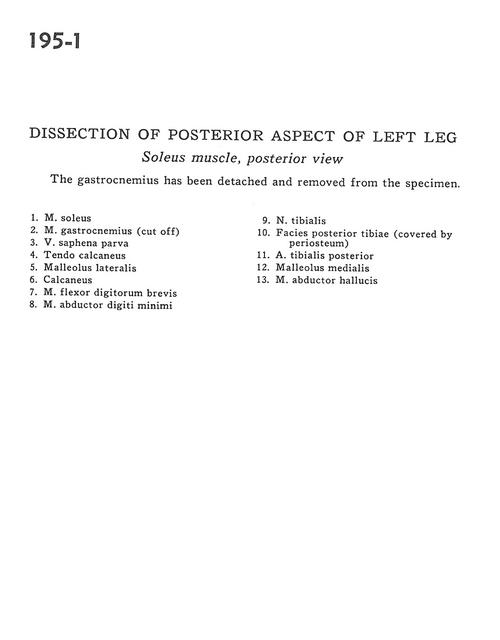
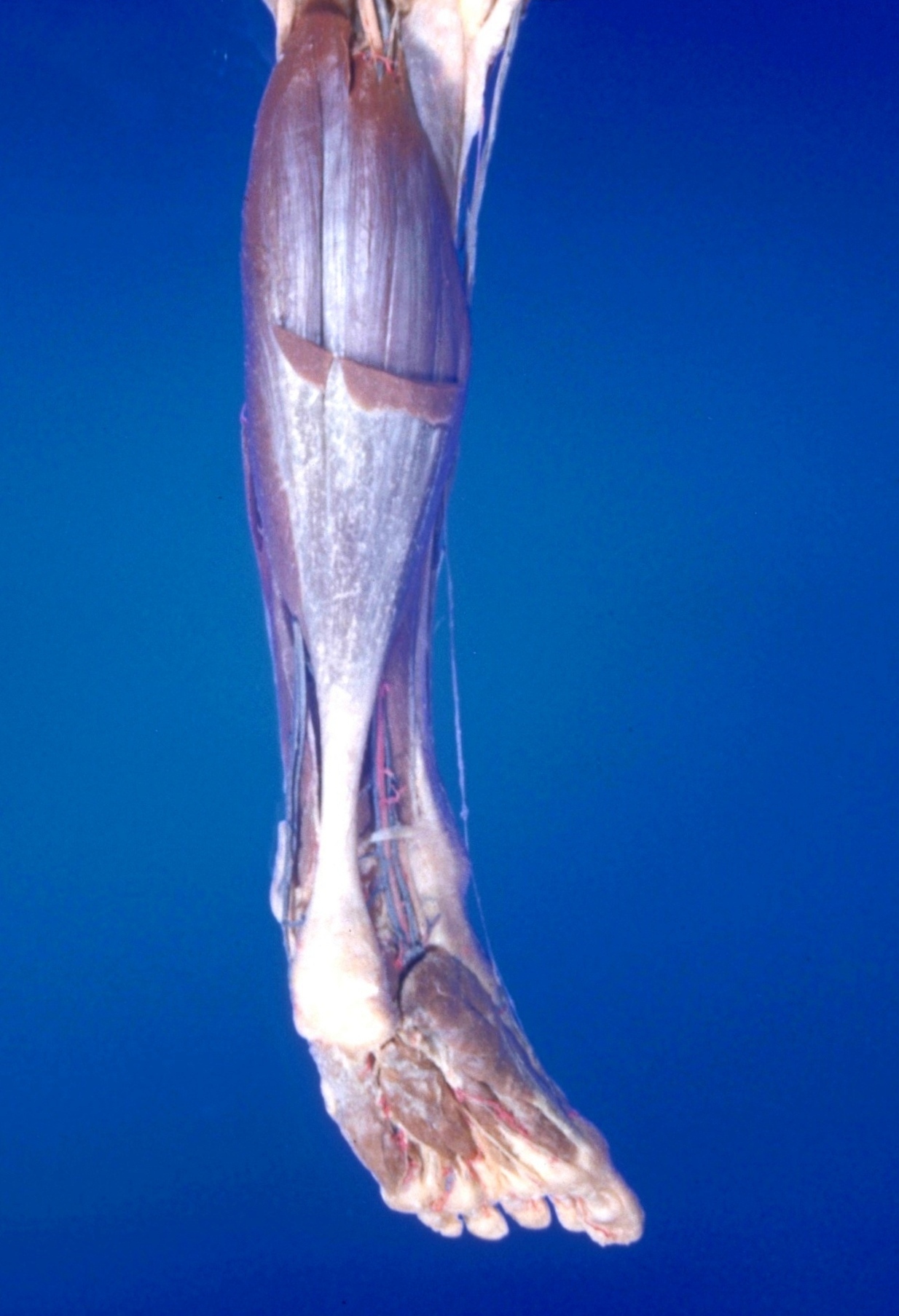
Dissection of posterior aspect of left leg

Soleus muscle, posterior view

Stanford holds the copyright to the David L. Bassett anatomical images and has assigned Creative Commons license Attribution-Share Alike 4.0 International to all of the images.
For additional information regarding use and permissions, please contact Dr. Drew Bourn at dbourn@stanford.edu.

Image #195-1
Bassett ImageDiagram Image
Legend Image
Dissection of posterior aspect of left leg
Soleus muscle, posterior view
The gastrocnemius has been detached and removed from teh specimen.
1 . Soleus muscle
2 . Gastrocnemius muscle (cut off)
3 . Lesser saphenous vein
4 . Tendo calcaneus
5 . Lateral malleolus
6 . Calcaneus
7 . Flexor digitorum brevis muscle
8 . Abductor digiti minimi muscle
9 . Tibial nerve
10 . Posterior surface of tibia (covered by periosteum)
11 . Posterior tibial artery
12 . Medial malleolus
13 . Abductor hallucis muscle