X CLOSE

Radiography

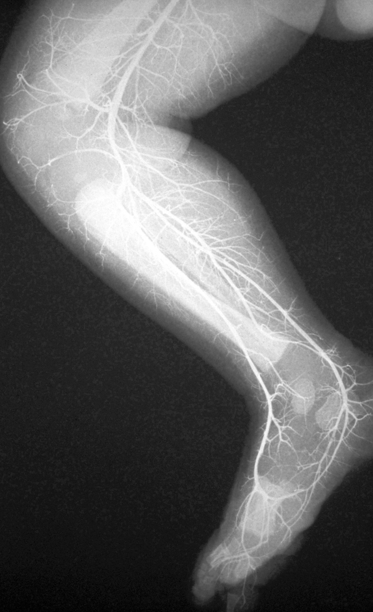
Angiogram, right lower limb of newborn infant

Stanford holds the copyright to the David L. Bassett anatomical images and has assigned Creative Commons license Attribution-Share Alike 4.0 International to all of the images.
For additional information regarding use and permissions, please contact Dr. Drew Bourn at dbourn@stanford.edu.

Image #179-2
Bassett ImageDiagram Image
Legend Image
Radiography
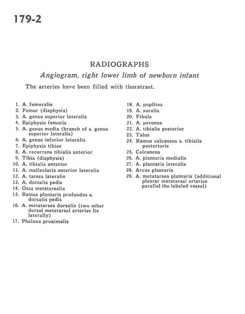
Angiogram, right lower limb of newborn infant
The arteries have been filled with thoratrast.
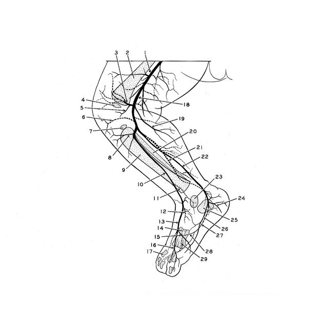
1 . Femoral artery
2 . Femur (diaphysis)
3 . Lateral superior genicular artery
4 . Femur (epiphysis)
5 . Medial genicular artery (branch of lateral superior genicular artery)
6 . Lateral inferior genicular artery
7 . Tibia (epiphysis)
8 . Anterior tibial recurrent artery
9 . Tibia (diaphysis)
10 . Anterior tibial artery
11 . Anterior lateral malleolar artery
12 . Lateral tarsal artery
13 . Dorsalis pedis artery
14 . Metatarsal bone
15 . Deep plantar branch of dorsalis pedis artery
16 . Dorsal metatarsal artery (two other dorsal metatarsal arteries lie laterally)
17 . Proximal phalanx
18 . Popliteal artery
19 . Sural artery
20 . Fibula
21 . Peroneal artery
22 . Posterior tibial artery
23 . Talus
24 . Ramus calcaneus branch of the posterior tibial artery
25 . Calcaneus
26 . Medial plantar artery
27 . Lateral plantar artery
28 . Arcus plantaris
29 . Plantar metatarsal artery (additional plantar metatarsal arteries parallel the labeled vessel)