X CLOSE

Dissection of female pelvis from a lateral approach

Blood vessels and nerves of uterus, vagina and bladder, anterior view

Stanford holds the copyright to the David L. Bassett anatomical images and has assigned Creative Commons license Attribution-Share Alike 4.0 International to all of the images.
For additional information regarding use and permissions, please contact Dr. Drew Bourn at dbourn@stanford.edu.

Image #163-1
Bassett ImageDiagram Image
Legend Image
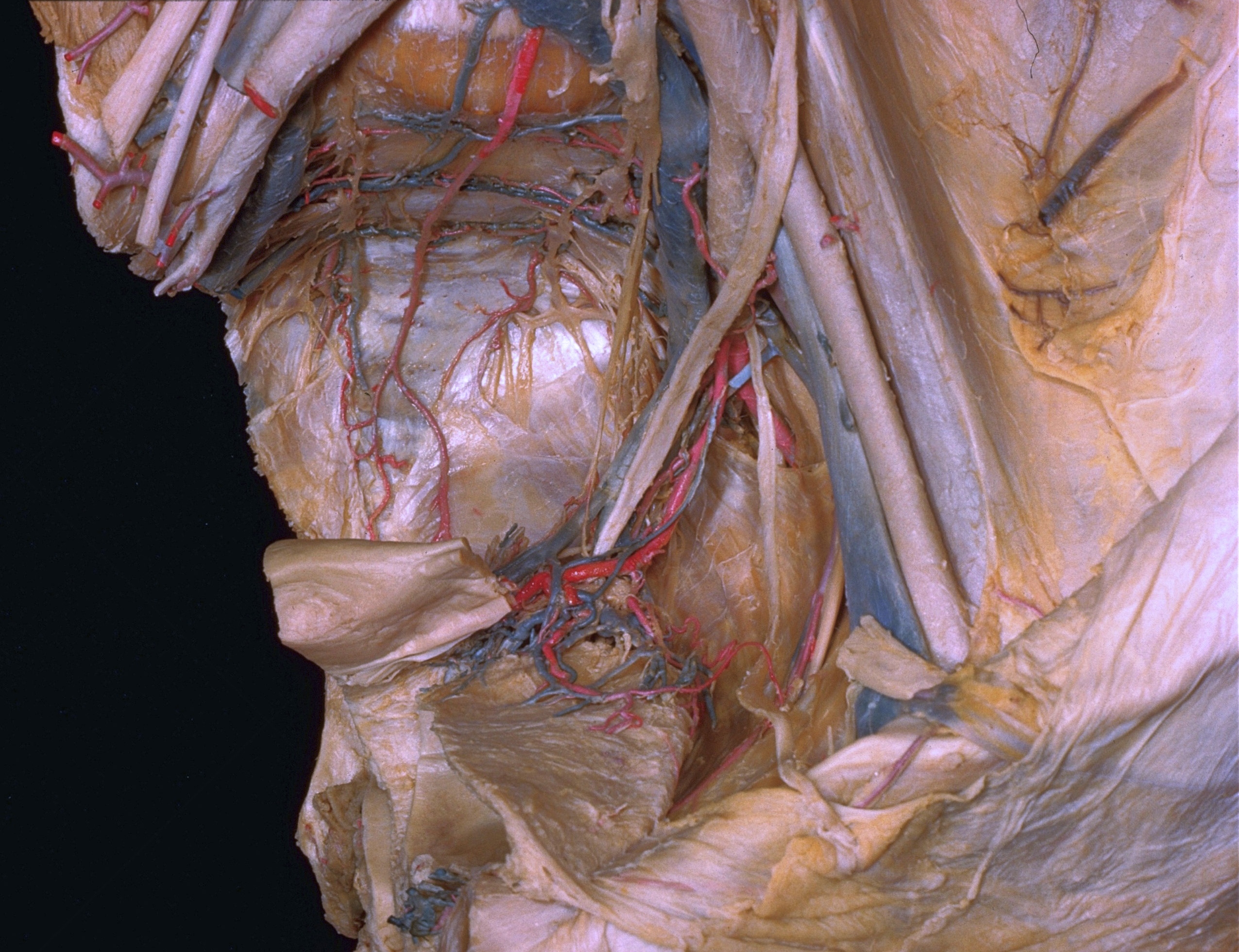
Dissection of female pelvis from a lateral approach
Blood vessels and nerves of uterus, vagina and bladder, anterior view
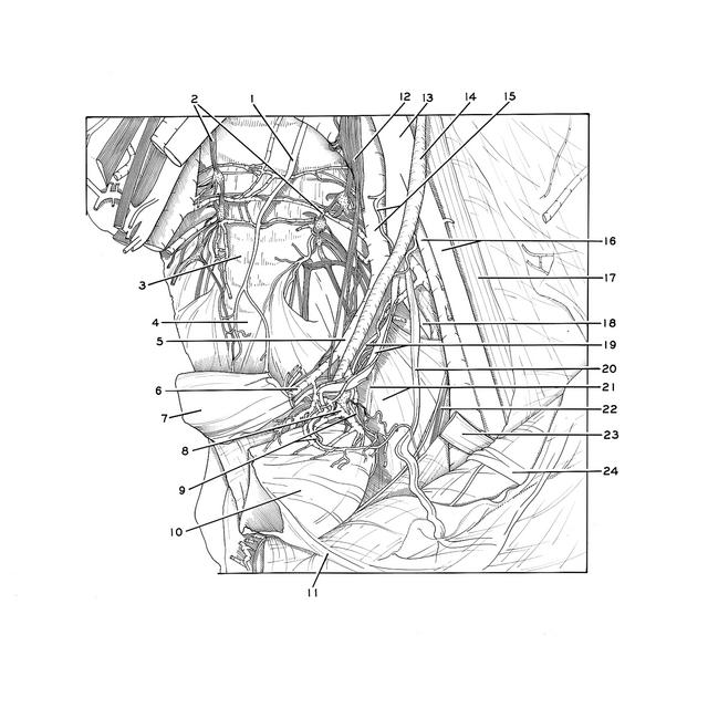
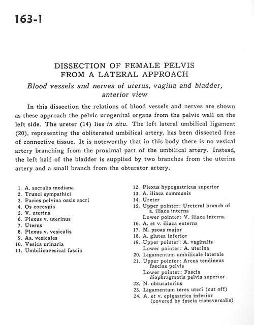
In this dissection the relations of blood vessels and nerves are shown as these approach the pelvic urogenital organs from the pelvic wall on the left side. The ureter (14) lies in situ. The left lateral umbilical ligament (20), representing the obliterated umbilical artery, has been dissected free of connective tissue. It is noteworthy that in this body there is no vesical artery branching from the proximal part of the umbilical artery. Instead, the left half of the bladder is supplied by two branches from the uterine artery and a small branch from the obturator artery.
1 . Middle sacral artery
2 . Sympathetic trunks
3 . Pelvic surface of sacrum
4 . Coccyx
5 . Uterine vein
6 . Uterine venous plexus
7 . Uterus
8 . Vesical venous plexus
9 . Vesical arteries
10 . Urinary bladder
11 . Umbilicovesical fascia
12 . Superior hypogastric plexus
13 . Common iliac artery
14 . Ureter
15 . Upper pointer: Ureteral branch of internal iliac artery Lower pointer: Internal iliac vein
16 . External iliac artery and vein
17 . Psoas major muscle
18 . Inferior gluteal artery
19 . Upper pointer: Vaginal artery Lower pointer: Uterine artery
20 . Lateral umbilical ligament
21 . Upper pointer: Tendinous arch of pelvic fascia Lower pointer: Superior fascia of pelvic diaphragm
22 . Obturator nerve
23 . Ligamentum teres (of uterus) (cut off)
24 . Inferior epigastric artery and vein (covered by transversalis fascia)