X CLOSE

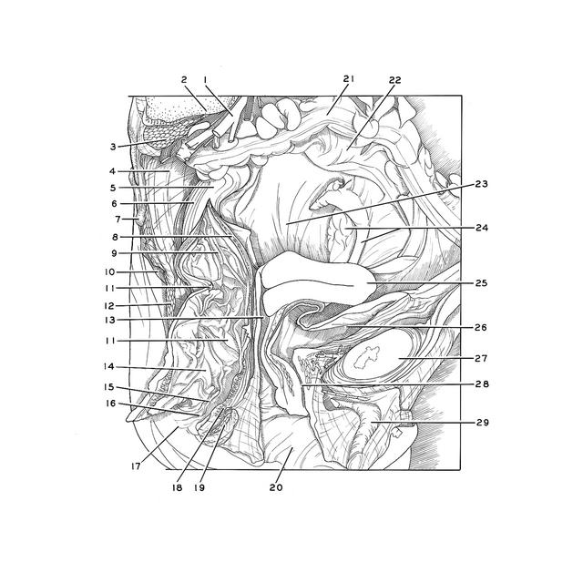
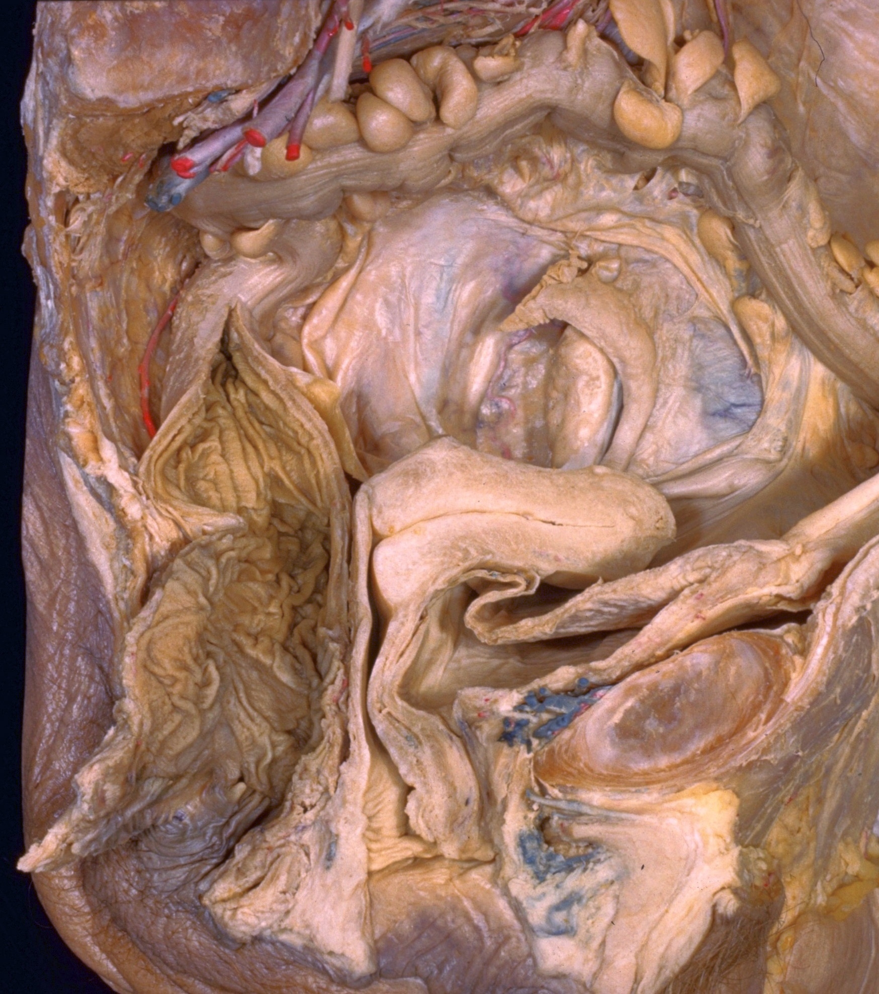
Dissection of female pelvis from a lateral approach

Interior of rectum and anal canal, general view

Stanford holds the copyright to the David L. Bassett anatomical images and has assigned Creative Commons license Attribution-Share Alike 4.0 International to all of the images.
For additional information regarding use and permissions, please contact Dr. Drew Bourn at dbourn@stanford.edu.

Image #161-7
Bassett ImageDiagram Image
Legend Image
Dissection of female pelvis from a lateral approach
Interior of rectum and anal canal, general view
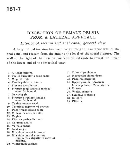
A longitudinal incision has been made through the anterior wall of the anal canal and rectum from the anus to the level of the sacral flexure. The wall to the right of the incision has been pulled aside to reveal the lumen of the lower end of the intestinal tract.
1 . Internal iliac artery
2 . Articular surface of sacrum
3 . Piriform muscle
4 . Parietal pelvic fascia
5 . Sacral flexure of rectum
6 . Longitudinal layer of muscular tunic of rectum
7 . Coccyx
8 . Circular layer of muscular tunic of rectum
9 . Mucosal tunic of rectum
10 . Terminal segment of coccyx
11 . Transverse fold of rectum
12 . Levator ani muscle (cut off)
13 . Vagina
14 . Perineal flexure of rectum
15 . Anal column
16 . Anal valve
17 . Anal bend
18 . Internal anal sphincter muscle
19 . External anal sphincter muscle (cut across, slightly to right of midline)
20 . Vestibule of vagina
21 . Sigmoid colon
22 . Mesosigmoid colon
23 . Rectouterine fold
24 . Upper pointer: Ovary Lower pointer: Uterine tube
25 . Uterus
26 . Urinary bladder
27 . Pubic symphysis
28 . Urethra
29 . Clitoris