Dissection of female pelvis from a lateral approach
Pubis, ischium and external genitalia, close-up view of right lateral aspect
For permissions information regarding the use of these images,
please contact Drew Bourn at dbourn@stanford.edu
Image #158-3
Dissection of female pelvis from a lateral approach
Pubis, ischium and external genitalia, close-up view of right lateral aspect
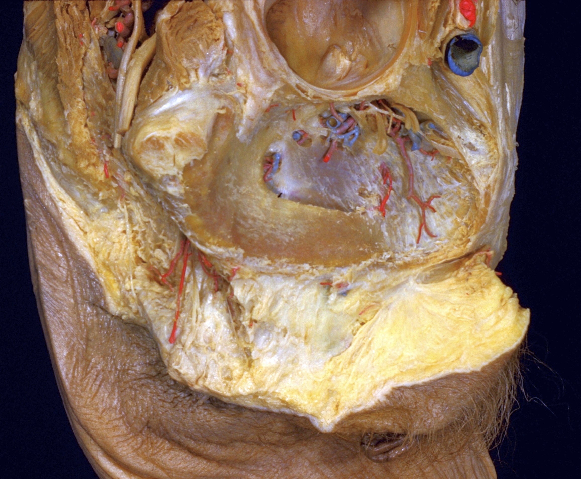
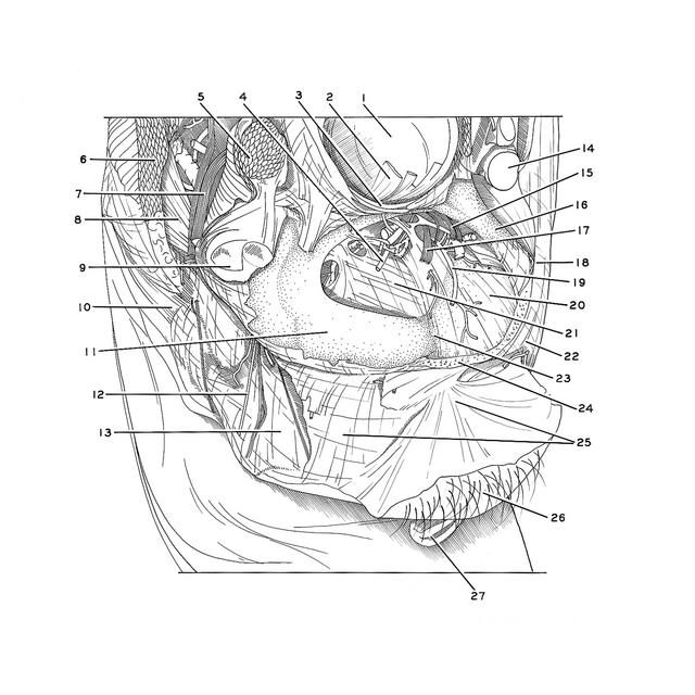
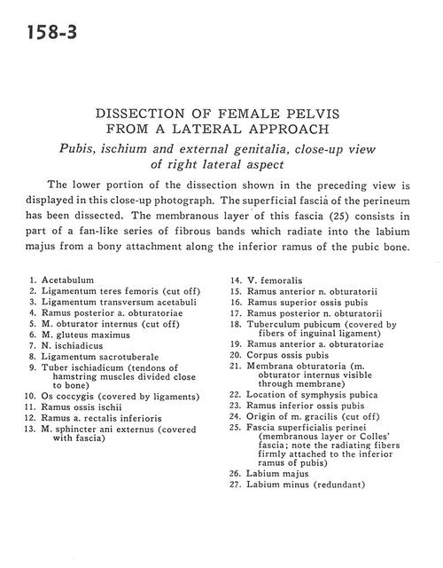
The lower portion of the dissection shown in the preceding view is displayed in this close-up photograph. The superficial fascia of the perineum has been dissected. The membranous layer of the fascia (25) consists in part of a fan-like series of fibrous bands which radiate into the labium majus from a bony attachment along the inferior ramus of the pubic bone.
1
.
Acetabulum
2
.
Ligamentum teres femoris (cut off)
3
.
Ligamentum transversum of acetabulum
4
.
Posterior branch of obturator artery
5
.
Obturator internus muscle (cut off)
6
.
Gluteus maximus muscle
7
.
Sciatic nerve
8
.
Sacrotuberous ligament
9
.
Ischial tuberosity (tendons of hamstring muscles divided close to bone)
10
.
Coccyx (covered by ligaments)
11
.
Ramus of ischium
12
.
Branch of inferior rectal artery
13
.
External anal sphincter muscle (covered with fascia)
14
.
Femoral vein
15
.
Anterior branch of obturator nerve
16
.
Superior pubic ramus
17
.
Posterior branch of obturator nerve
18
.
Pubic tubercle (covered by fibers of inguinal ligament)
19
.
Anterior branch of obturator artery
20
.
Body of pubic bone
21
.
Obturator membrane (obturator internus muscle visible through membrane)
22
.
Location of pubic symphysis
23
.
Inferior pubic ramus
24
.
Origin of gracilis muscle (cut off)
25
.
Superficial perineal fascia (membranous layer or Colles' fascia; note the radiating fibers firmly attached to the inferior ramus of pubis)