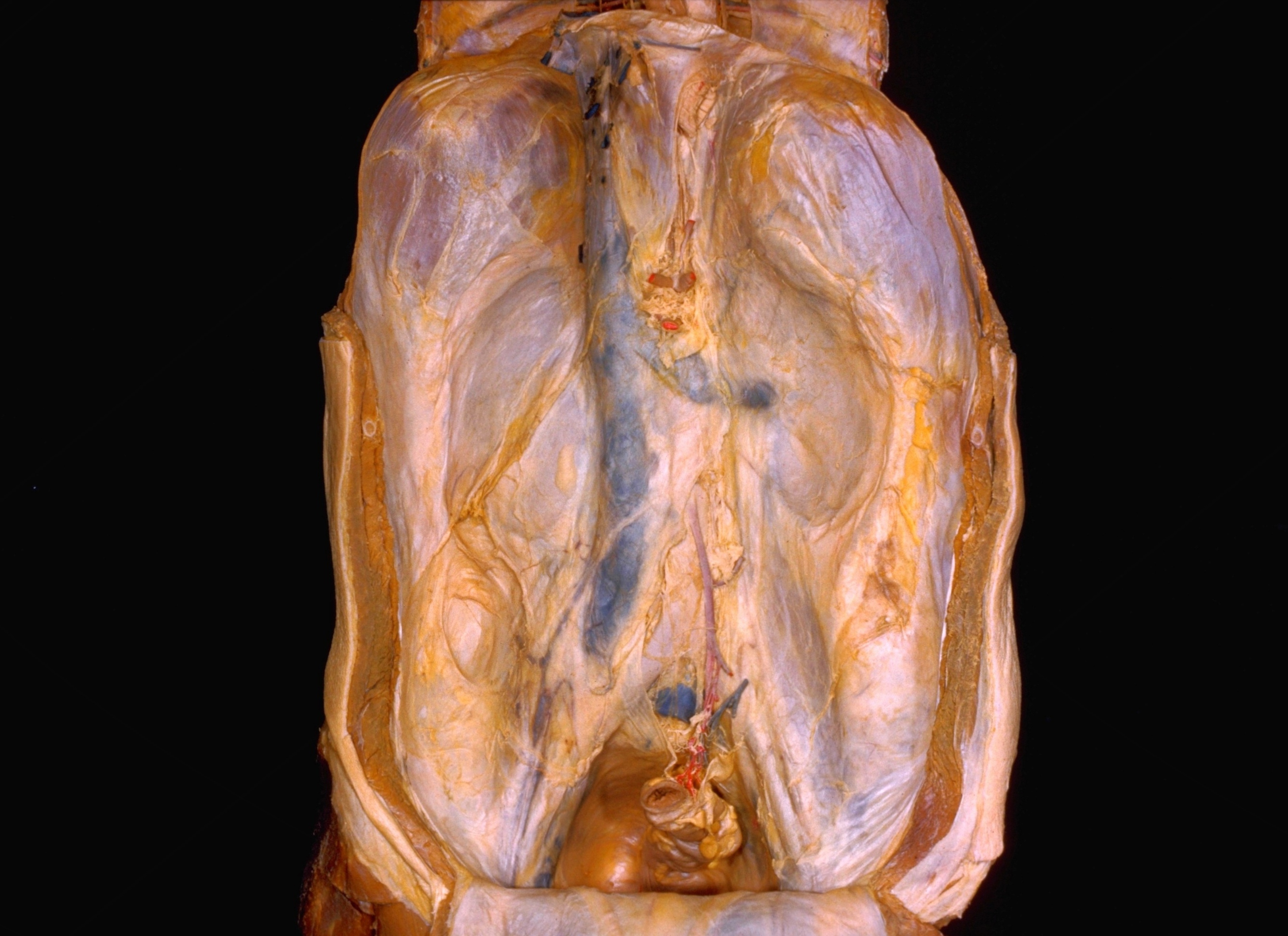
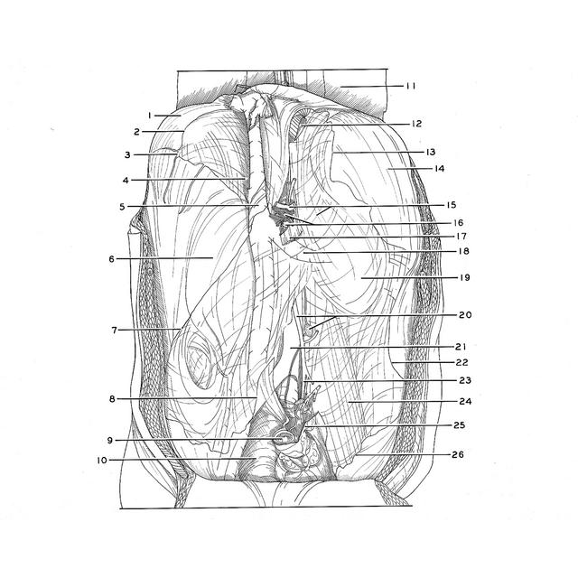
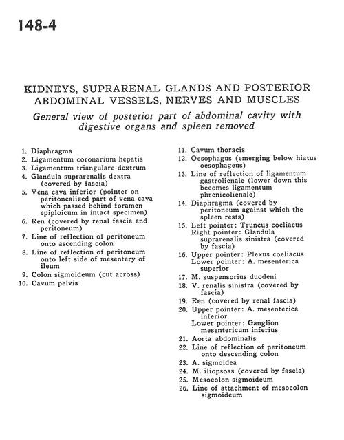
Kidneys, suprarenal glands and posterior abdominal vessels, nerves and muscles
General view of posterior part of abdominal cavity with digestive organs and spleen removed
Stanford holds the copyright to the David L. Bassett anatomical images and has assigned
Creative Commons license Attribution-Share Alike 4.0 International to all of the images.
For additional information regarding use and permissions,
please contact Dr. Drew Bourn at dbourn@stanford.edu.
Image #148-4
 |  | ||||||||||||||||||||||||||||||||||||||||||||||||||||||||
 |
|