Exploration of liver, gall bladder, pancreas, duodenum and spleen
Posteroinferior aspect of liver
Stanford holds the copyright to the David L. Bassett anatomical images and has assigned
Creative Commons license Attribution-Share Alike 4.0 International to all of the images.
For additional information regarding use and permissions,
please contact Dr. Drew Bourn at dbourn@stanford.edu.
Image #144-7
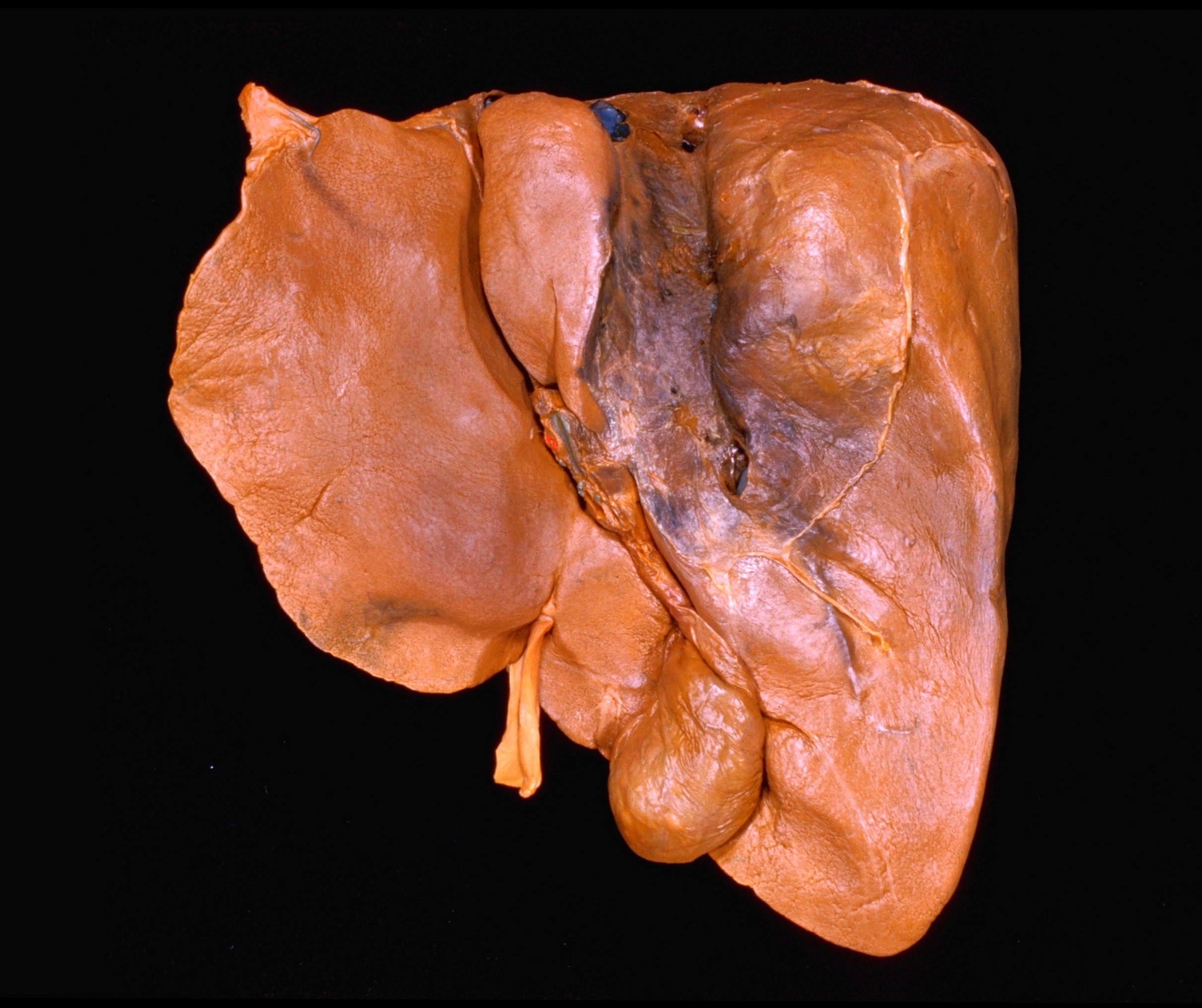 | 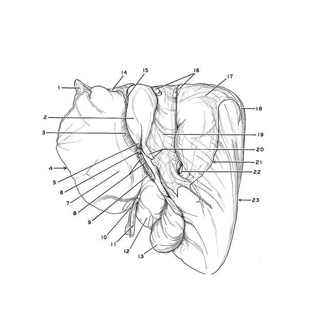 |
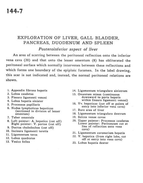 | | Exploration of liver, gall bladder, pancreas, duodenum and spleen | | Posteroinferior aspect of liver | | An area of scarring between the peritoneal reflection onto the inferior vena cava (20) and that onto the lesser omentum (8) has obliterated the peritoneal surface which normally intervenes between these reflections and which forms one boundary on the epiploic foramen. In the label drawing, this scar is not indicated and, instead, the normal peritoneal relations are shown. | | 1
.
| Left triangular ligament of liver | | 2
.
| Caudate lobe | | 3
.
| Fissure of ligamentum venosum | | 4
.
| Left lobe of liver | | 5
.
| Papillary process | | 6
.
| Hepatic lymph node (sectioned in division of lesser omentum) | | 7
.
| Omental tubercle | | 8
.
| Left pointer: Hepatic artery (cut off) Right pointer: Portal vein (cut off) | | 9
.
| Common bile duct (cut off) | | 10
.
| Notch for ligamentum teres | | 11
.
| Ligamentum teres | | 12
.
| Quadrate lobe | | 13
.
| Gallbladder | | 14
.
| Left triangular ligament | | 15
.
| Lesser omentum (continuous downward to porta hepatis within fissure of ligamentum venosum) | | 16
.
| Hepatic veins (cut off at points of entry into inferior vena cava) | | 17
.
| Bare area of liver | | 18
.
| Right triangular ligament | | 19
.
| Sulcus of vena cava | | 20
.
| Upper pointer: Caudate process Lower pointer: Peritoneum cut at line of reflection onto vena cava) | | 21
.
| Coronary ligament of liver | | 22
.
| Hepatic vein (from right lobe, cut off at entry into vena cava) | | 23
.
| Right lobe of liver |
|
|


