X CLOSE

Dissection of diaphragm

Sternal attachment of diaphragm.

Stanford holds the copyright to the David L. Bassett anatomical images and has assigned Creative Commons license Attribution-Share Alike 4.0 International to all of the images.
For additional information regarding use and permissions, please contact Dr. Drew Bourn at dbourn@stanford.edu.

Image #130-5
Bassett ImageDiagram Image
Legend Image
Dissection of diaphragm
Sternal attachment of diaphragm.
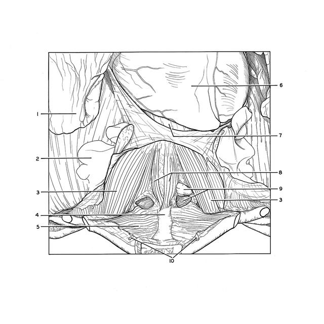
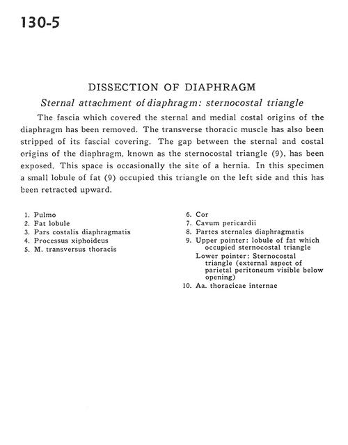
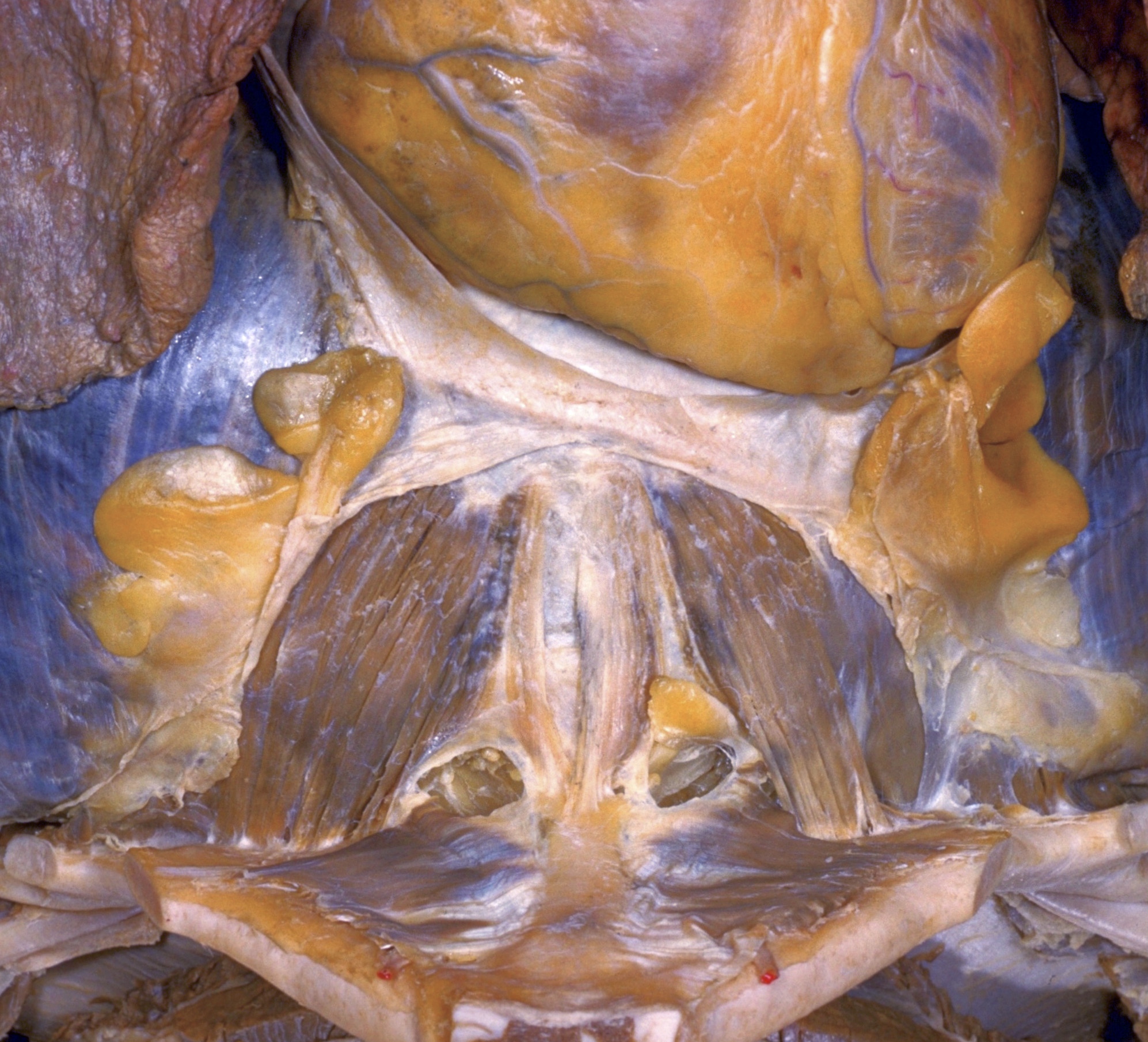
The fascia which covered the sternal and medial costal origins of the diaphragm has been removed. The transverse thoracic muscle has also been stripped of its fascial covering. The gap between the sternal and costal origins of the diaphragm, known as the sternocostal triangle (9), has been exposed. This space is occasionally the site of a hernia. In this specimen a small lobule of fat (9) occupied this triangle on the left side and this has been retracted upward.
1 . Lung
2 . Fat lobule
3 . Costal part of diaphragm
4 . Xiphoid process
5 . Transversus thoracis muscle
6 . Heart
7 . Pericardial cavity
8 . Sternal part of diaphragm
9 . Upper pointer: Lobule of fat which occupied sternocostal triangle Lower pointer: Sternocostal triangle (external aspect of parietal peritoneum visible below opening)
10 . Internal thoracic artery