X CLOSE

Detailed dissection of heart

Coronary angiogram

Stanford holds the copyright to the David L. Bassett anatomical images and has assigned Creative Commons license Attribution-Share Alike 4.0 International to all of the images.
For additional information regarding use and permissions, please contact Dr. Drew Bourn at dbourn@stanford.edu.

Image #119-7
Bassett ImageDiagram Image
Legend Image
Detailed dissection of heart
Coronary angiogram
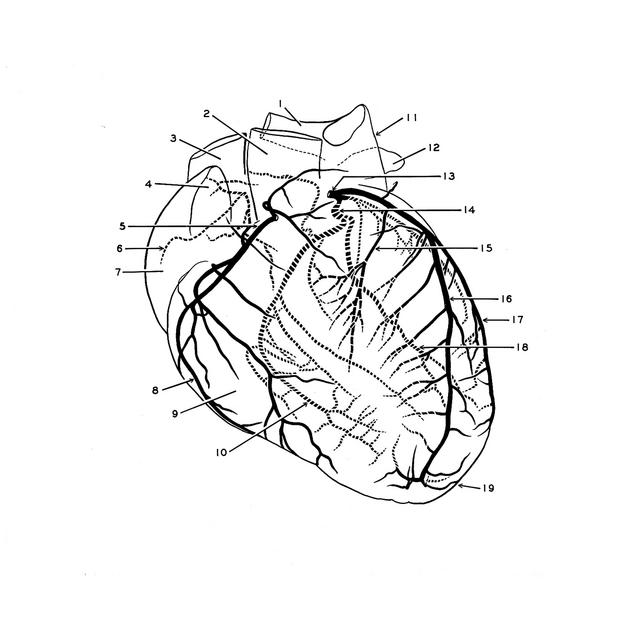
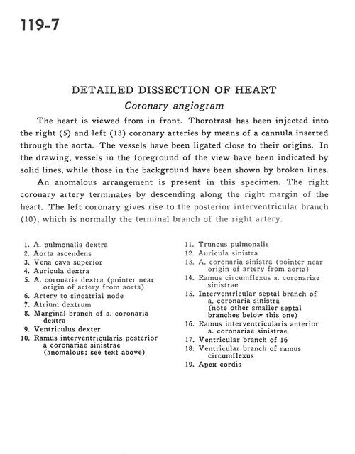
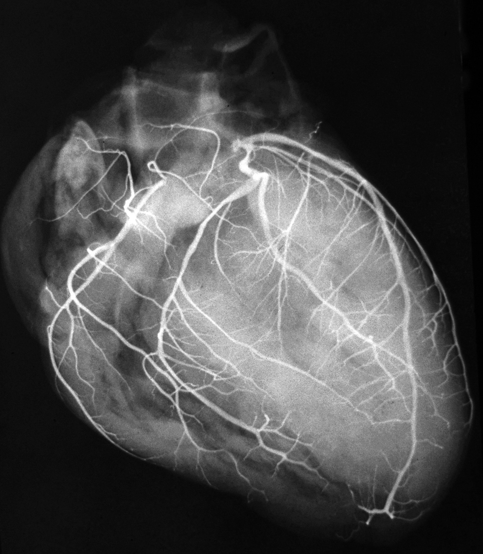
The heart is viewed from in front. Thorotrast has been injected into the right (5) and left (13) coronary arteries by means of a cannula inserted through the aorta. The vessels have been ligated close to their origins. In the drawing, vessels in the foreground of the view have been indicated by solid lines, while those in the background have been shown by broken lines.
1 . Right pulmonary artery
2 . Ascending aorta
3 . Superior vena cava
4 . Right auricle
5 . Right coronary artery (pointer near origin of artery from aorta)
6 . Artery to sinoatrial node
7 . Right atrium
8 . Marginal branch of right coronary artery
9 . Right ventricle
10 . Posterior interventricular branch of left coronary artery (anomalous see text above)
11 . Pulmonary trunk
12 . Left auricle
13 . Left coronary artery (pointer near origin of artery from aorta)
14 . Circumflex branch of left coronary artery
15 . Interventricular septal branch of left coronary artery (note other smaller septal branches below this one)
16 . Anterior interventricular branch of left coronary artery
17 . Ventricular branch of 16
18 . Ventricular branch of circumflex branch
19 . Apex of heart