X CLOSE

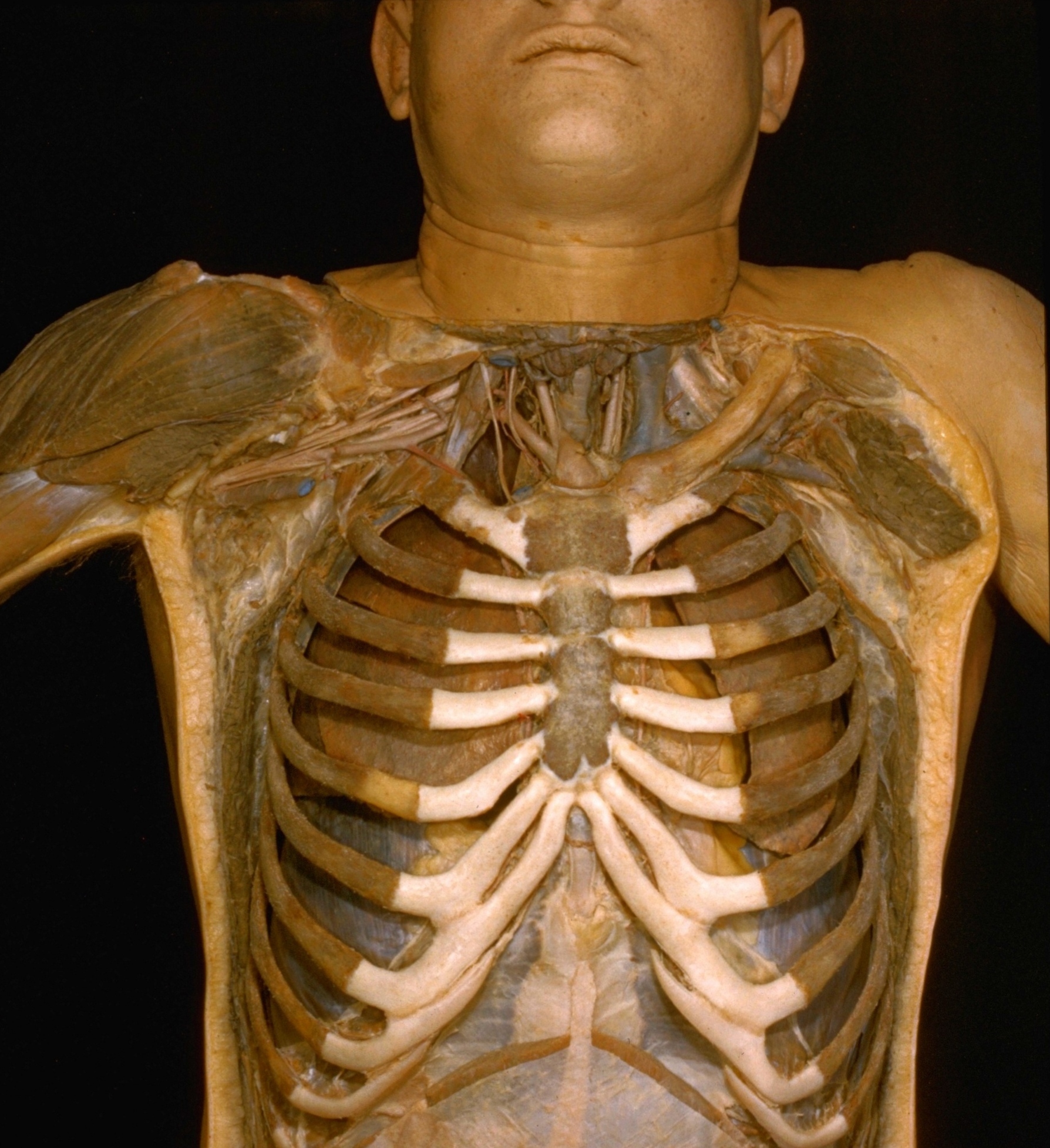
Thoracic viscera in situ

Anterior view of thoracic contents, rib cage intact

Stanford holds the copyright to the David L. Bassett anatomical images and has assigned Creative Commons license Attribution-Share Alike 4.0 International to all of the images.
For additional information regarding use and permissions, please contact Dr. Drew Bourn at dbourn@stanford.edu.

Image #116-2
Bassett ImageDiagram Image
Legend Image
Thoracic viscera in situ
Anterior view of thoracic contents, rib cage intact
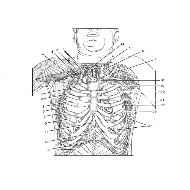
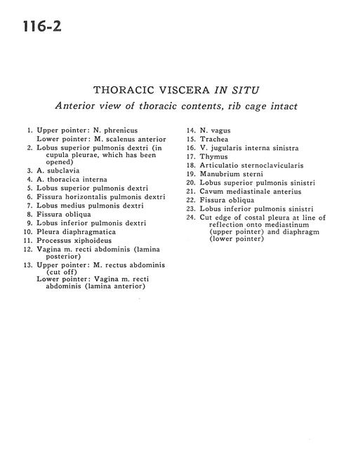
1 . Upper pointer: Phrenic nerve Lower pointer: Anterior scalene muscle
2 . Upper lobe right lung (in cupula pleurae, which has been opened)
3 . Subclavian artery
4 . Internal thoracic artery
5 . Upper lobe right lung
6 . Horizontal fissure right lung
7 . Middle lobe right lung
8 . Oblique fissure
9 . Inferior lobe right lung
10 . Diaphragmatic pleura
11 . Xiphoid process
12 . Sheath rectus abdominis muscle (posterior surface)
13 . Upper pointer: Rectus abdominis muscle (cut off) Lower pointer: Sheath of rectus abdominis muscle (anterior surface)
14 . Vagus nerve
15 . Trachea
16 . Left internal jugular vein
17 . Thymus
18 . Sternoclavicular joint
19 . Manubrium of sternum
20 . Upper lobe left lung
21 . Anterior mediastinum
22 . Oblique fissure
23 . Lower lobe left lung
24 . Cut edge of costal pleura at line of reflection onto mediastinum (upper pointer) and diaphragm (lower pointer)