| 1
.
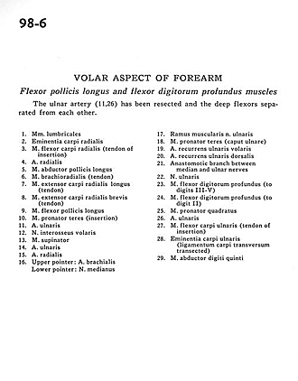
| Lumbrical muscles |
| 2
.
| Radial carpal eminence |
| 3
.
| Flexor carpi radialis muscle (tendon of insertion) |
| 4
.
| Radial artery |
| 5
.
| Abductor pollicis longus muscle |
| 6
.
| Brachioradialis muscle (tendon) |
| 7
.
| Extensor carpi radialis longus muscle (tendon) |
| 8
.
| Extensor carpi radialis brevis muscle (tendon) |
| 9
.
| Flexor pollicis longus muscle |
| 10
.
| Pronator teres muscle (insertion) |
| 11
.
| Ulnar artery |
| 12
.
| Anterior interosseous nerve |
| 13
.
| Supinator muscle |
| 14
.
| Ulnar artery |
| 15
.
| Radial artery |
| 16
.
| Upper pointer: Brachial artery Lower pointer: Median nerve |
| 17
.
| Muscular branch of ulnar nerve |
| 18
.
| Pronator teres muscle (ulnar head) |
| 19
.
| Anterior recurrent ulnar artery |
| 20
.
| Dorsal recurrent ulnar artery |
| 21
.
| Anastomotic branch between median and ulnar nerves |
| 22
.
| Ulnar nerve |
| 23
.
| Flexor digitorum profundus muscle (to digits III-V) |
| 24
.
| Flexor digitorum profundus muscle (to digit II) |
| 25
.
| Pronator quadratus muscle |
| 26
.
| Ulnar artery |
| 27
.
| Flexor carpi ulnaris muscle (tendon of insertion) |
| 28
.
| Ulnar carpal eminence (ligamentum carpi transversum transected) |
| 29
.
| Abductor digiti minimi muscle |