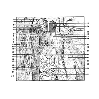
| 1
.
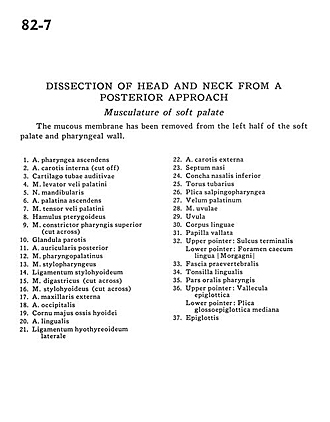
| Ascending pharyngeal artery |
| 2
.
| Internal carotid artery (cut off) |
| 3
.
| Cartilaginous auditory tube |
| 4
.
| Levator veli palatini muscle |
| 5
.
| Mandibular nerve |
| 6
.
| Ascending palatine artery |
| 7
.
| Tensor veli palatini muscle |
| 8
.
| Pterygoid hamulus |
| 9
.
| Superior pharyngeal constrictor muscle (cut across) |
| 10
.
| Parotid gland |
| 11
.
| Posterior auricular artery |
| 12
.
| Palatopharyngeus muscle |
| 13
.
| Stylopharyngeus muscle |
| 14
.
| Stylohyoid ligament |
| 15
.
| Stylohyoid muscle (cut across) |
| 16
.
| Digastric muscle (cut across) |
| 17
.
| External maxillary artery |
| 18
.
| Occipital artery |
| 19
.
| Greater horn hyoid bone |
| 20
.
| Lingual artery |
| 21
.
| Lateral thyrohyoid ligament |
| 22
.
| External carotid artery |
| 23
.
| Nasal septum |
| 24
.
| Inferior nasal concha |
| 25
.
| Torus tubarius |
| 26
.
| Salpingopharyngeal fold |
| 27
.
| Velum of palate |
| 28
.
| Uvular muscle |
| 29
.
| Uvula |
| 30
.
| Body of tongue |
| 31
.
| Vallate papilla |
| 32
.
| Upper pointer: Sulcus terminalis Lower pointer: Foramen caecum |
| 33
.
| Prevertebral fascia |
| 34
.
| Lingual tonsil |
| 35
.
| Oral part pharynx |
| 36
.
| Upper pointer: Epiglottic vallecula Lower pointer: Median glossoepiglottic fold |
| 37
.
| Epiglottis |


