Bassett Collection of Stereoscopic Images of Human Anatomy
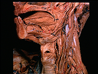
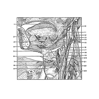
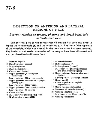
Dissection of anterior and lateral regions of neck
Larynx; relation to tongue, pharynx and hyoid bone, left anterolateral view
Image #77-6
KEYWORDS: Face, Mouth, Muscles and tendons, Pharynx, Throat.
Creative Commons
Stanford holds the copyright to the David L. Bassett anatomical images and has assigned Creative Commons license Attribution-Share Alike 4.0 International to all of the images.

For additional information regarding use and permissions, please contact Dr. Drew Bourn at dbourn@stanford.edu.
|
| ||||||||||||||||||||||||||||||||||||||||||||||||||
|
|