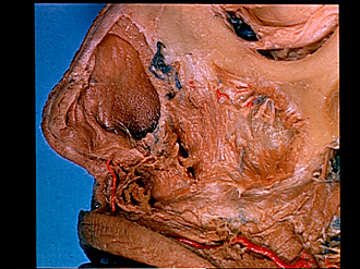
| 1
.
| Nasal bone |
| 2
.
| External nasal branch of anterior ethmoidal nerve |
| 3
.
| Lateral nasal cartilage (cut away) |
| 4
.
| Nasal septum |
| 5
.
| Greater alar cartilage (lateral crus cut away) |
| 6
.
| Nasal mucosal membrane |
| 7
.
| Apex nasi |
| 8
.
| Hair |
| 9
.
| Vestibule (of nose) |
| 10
.
| Cut edge of skin of mobile nasal septum |
| 11
.
| Artery of nasal septum |
| 12
.
| Depressor septi nasi muscle (lateral fibers form alar part nasalis muscle, not a separate muscle in this case) |
| 13
.
| Superior labial branches infraorbital nerve (cut ofl) |
| 14
.
| Labial gland |
| 15
.
| Orbicularis oris muscle |
| 16
.
| Frontal process of maxilla |
| 17
.
| Inferior oblique muscle |
| 18
.
| Infraorbital margin |
| 19
.
| Zygomaticomaxillary suture |
| 20
.
| Infraorbital head of levator labii superioris muscle |
| 21
.
| Branches of infraorbital nerve emerging from infraorbital foramen |
| 22
.
| Transverse part of nasalis muscle (cut off) |
| 23
.
| Canine fossa |
| 24
.
| Remnant of angular head of levator labii superioris muscle |
| 25
.
| Internal nasal branch infraorbital nerve (cut off) |
| 26
.
| Depressor anguli oris muscle (cut off) |
| 27
.
| Buccinator muscle |
| 28
.
| Superior labial artery |


