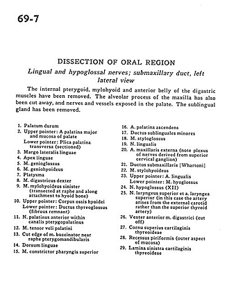
| 1
.
| Hard palate |
| 2
.
| Upper pointer: Greater palatine artery and mucosa of palate Lower pointer: Transverse palatine fold (sectioned) |
| 3
.
| Lateral lingual margin |
| 4
.
| Apex of tongue |
| 5
.
| Genioglossus muscle |
| 6
.
| Geniohyoid muscle |
| 7
.
| Platysma |
| 8
.
| Digastric muscle right |
| 9
.
| Mylohyoid muscle left (transected at raphe and along attachment to hyoid bone) |
| 10
.
| Upper pointer: Body of hyoid bone Lower pointer: Thyroglossal duct (fibrous remnant) |
| 11
.
| Anterior palatine nerve within pterygopalatine canal |
| 12
.
| Tensor veli palatini muscle |
| 13
.
| Cut edge of buccinator muscle near pterygomandibular raphe |
| 14
.
| Dorsum of tongue |
| 15
.
| Superior pharyngeal constrictor muscle |
| 16
.
| Ascending palatine artery |
| 17
.
| Minor sublingual duct |
| 18
.
| Styloglossus muscle |
| 19
.
| Lingual nerve |
| 20
.
| External maxillary artery (note plexus of nerves derived from superior cervical ganglion) |
| 21
.
| Submandibular duct |
| 22
.
| Stylohyoid muscle |
| 23
.
| Upper pointer: Lingual artery Lower pointer: Hyoglossus muscle |
| 24
.
| Hypoglossal nerve (XII) |
| 25
.
| Superior laryngeal nerve and superior laryngeal artery (in this case the artery arises from the external carotid rather than the superior thyroid artery) |
| 26
.
| Anterior belly digastric muscle (cut off) |
| 27
.
| Superior horn thyroid cartilage |
| 28
.
| Piriform recess (outer aspect of mucosa) |
| 29
.
| Left lamina thyroid cartilage |