Bassett Collection of Stereoscopic Images of Human Anatomy
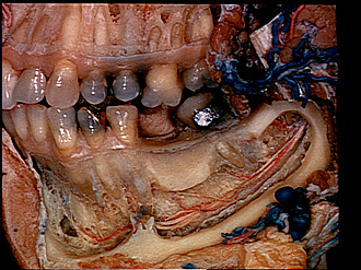
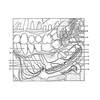
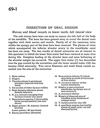
Dissection of oral region
Nerves and blood vessels to lower teeth, left lateral view
Image #69-1
KEYWORDS: Bones cartilage joints, Cheek, Face, Mouth, Peripheral nervous system, Vasculature.
Creative Commons
Stanford holds the copyright to the David L. Bassett anatomical images and has assigned Creative Commons license Attribution-Share Alike 4.0 International to all of the images.

For additional information regarding use and permissions, please contact Dr. Drew Bourn at dbourn@stanford.edu.
|
| ||||||||||||||||||||||||||||||||||||
|
|