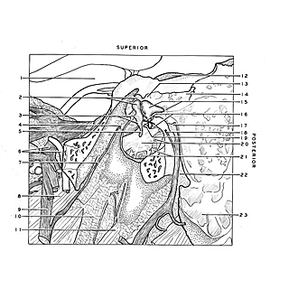
| 1
.
| Base of peduncle (in background) |
| 2
.
| Upper pointer: Superior ligament of malleus Lower pointer: Capitulum of malleus |
| 3
.
| Upper pointer: Process of anterior malleus (covered by anterior malleolar plica) Lower pointer: Lateral process malleus (the superior recess, Prussak's space, is located just above this process and is closed laterally by a thin downward extension of squamous bone known as the scutum) |
| 4
.
| Petrotympanic fissure |
| 5
.
| Manubrium of malleus |
| 6
.
| Upper pointer: Mandibular nerve (V) Lower pointer: Middle meningeal artery |
| 7
.
| Chorda tympani (passing inferiorly to join lingual nerve) |
| 8
.
| Tensor veli palatini muscle |
| 9
.
| Lingual nerve |
| 10
.
| Alar fascia |
| 11
.
| Styloglossus muscle |
| 12
.
| Arcuate eminence |
| 13
.
| Epitympanic recess |
| 14
.
| Tympanic antrum |
| 15
.
| Left pointer: Body incus Right pointer: Posterior ligament of incus |
| 16
.
| Left pointer: Chorda tympani within tympanic cavity Right pointer: Tendon of stapedius muscle |
| 17
.
| Facial nerve (VII) within facial canal |
| 18
.
| Chorda tympani within canaliculus |
| 19
.
| Left pointer: Anterior crus of stapes Right pointer: Tympanic sinus |
| 20
.
| Upper pointer: Fenestrated cochlear fossa (separated from tympanic sinus above by promontory) Lower pointer: Promontory (tympanic plexus visible beneath mucosa) |
| 21
.
| Tympanic cells of jugular wall of tympanic cavity |
| 22
.
| Position of stylomastoid foramen (opened) |
| 23
.
| Mastoid process (dissected) |