Bassett Collection of Stereoscopic Images of Human Anatomy
Microradiograph of eye; central optic pathways and related structures
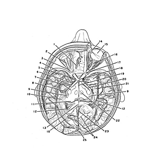
Optic pathways dissected in situ, viewed from above
Image #58A-2
KEYWORDS: Brain, Diencephalon, Eye, Face, Occipital lobe, Peripheral nervous system, Telencephalon.
Creative Commons
Stanford holds the copyright to the David L. Bassett anatomical images and has assigned Creative Commons license Attribution-Share Alike 4.0 International to all of the images.

For additional information regarding use and permissions, please contact Dr. Drew Bourn at dbourn@stanford.edu.

|
| ||||||||||||||||||||||||||||||||||||||||||||||||||||||||

|
|