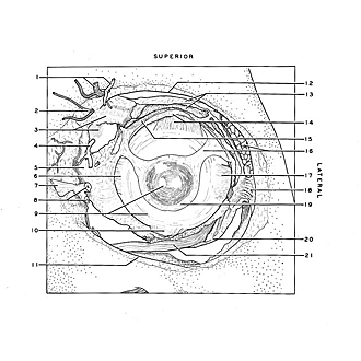
| 1
.
| Right pointer: Frontal artery Left pointer: Supratrochlear nerve |
| 2
.
| Infratrochlear nerve |
| 3
.
| Upper pointer: Superior oblique muscle (covered by fascia) Lower pointer: Superior oblique muscle (in background) |
| 4
.
| Middle palpebral artery (cut off) |
| 5
.
| Medial check ligament |
| 6
.
| Tendon of medial rectus muscle |
| 7
.
| Medial palpebral ligament |
| 8
.
| Lacrimal duct |
| 9
.
| Upper pointer: Cornea Lower pointer: Sclera |
| 10
.
| Tendon of inferior rectus muscle |
| 11
.
| Infraorbital margin |
| 12
.
| Supraorbital margin |
| 13
.
| Upper pointer: Fascia above Ievator palpebrae superioris muscle Lower pointer: Aponeurosis of levator palpebrae superioris (cut off) |
| 14
.
| Upper pointer: Fascia between levator palpebrae superioris and rectus superior muscles Lower pointer: Tendon of superior rectus muscle |
| 15
.
| Superior tarsalis muscle (muscle of Müller) (cut across) |
| 16
.
| Upper pointer: Lacrimal gland Lower pointer: Conjunctiva and bulbar fascia |
| 17
.
| Tendon of lateral rectus muscle |
| 18
.
| Lateral check ligament (attached to orbital tubercle of zygomatic bone) |
| 19
.
| Annulus conjunctivae overlapping limbus corneae |
| 20
.
| Inferior part of muscle fascia which forms suspensory ligament of eye |
| 21
.
| Inferior oblique muscle |