Bassett Collection of Stereoscopic Images of Human Anatomy
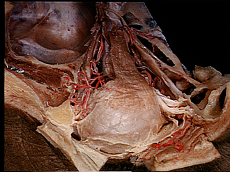
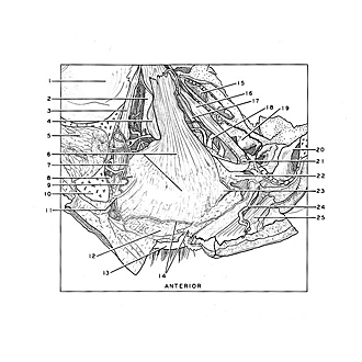
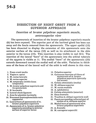
Dissection of right orbit from a superior approach
Insertion of levator palpebrae superioris muscle, anterosuperior view
Image #54-3
KEYWORDS: Eye, Face, Muscles and tendons.
Creative Commons
Stanford holds the copyright to the David L. Bassett anatomical images and has assigned Creative Commons license Attribution-Share Alike 4.0 International to all of the images.

For additional information regarding use and permissions, please contact Dr. Drew Bourn at dbourn@stanford.edu.
|
| ||||||||||||||||||||||||||||||||||||||||||||||||||||||
|
|