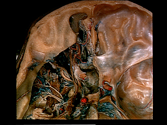
| 1
.
| Frontal sinus |
| 2
.
| Infundibulum leading into nasofrontal duct |
| 3
.
| Upper pointer: Frontal nerve Lower pointer: Levator palpebrae superioris |
| 4
.
| Posterior ethmoidal nerve (usually a branch of the ophthalmic nerve in this case the fibers accompany the trochlear nerve) |
| 5
.
| Posterior ethmoidal artery in posterior ethmoidal foramen |
| 6
.
| Lacrimal nerve and superior ophthalmic vein |
| 7
.
| Periorbita (area of fusion with common annular tendon) |
| 8
.
| Maxillary nerve (V2) |
| 9
.
| Trochlear nerve (IV) |
| 10
.
| Oculomotor nerve (III) |
| 11
.
| Abducens nerve (VI) |
| 12
.
| Vidian nerve of pterygoid canal (below floor of sphenoid sinus) |
| 13
.
| Semilunar ganglion (trigeminal) |
| 14
.
| Mandibular nerve (V3) |
| 15
.
| Cavernous nerve plexus |
| 16
.
| Ethmoidal cell (anterior) |
| 17
.
| Anterior ethmoidal nerve |
| 18
.
| Cribriform plate ethmoid bone |
| 19
.
| Anterior ethmoidal artery (in anterior ethmoidal foramen) |
| 20
.
| Ethmoidal cell (posterior) |
| 21
.
| Posterior ethmoidal vein |
| 22
.
| Upper pointer: Aperture of sphenoid sinus Lower pointer: Sphenoid sinus |
| 23
.
| Optic nerve (ll) |
| 24
.
| Internal carotid artery |
| 25
.
| Upper pointer: Circular sinus Lower pointer: Hypophysis (left half removed) |
| 26
.
| Sella turcica |
| 27
.
| Dorsum sellae |
| 28
.
| Basilar venous plexus |