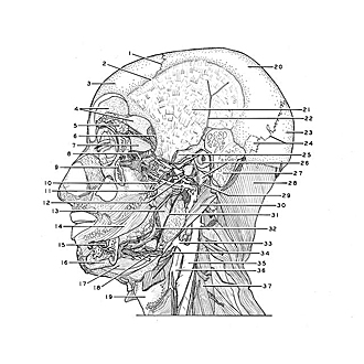
| 1
.
| Coronal suture |
| 2
.
| Superior temporal line |
| 3
.
| Frontal bone |
| 4
.
| Dura mater encephali (in anterior cranial fossa) |
| 5
.
| Superior lacrimal gland |
| 6
.
| Middle meningeal artery and dura mater encephali (in middle cranial fossa) |
| 7
.
| Lateral rectus muscle |
| 8
.
| Zygomatic nerve |
| 9
.
| Zygomatic bone (partially cut away) |
| 10
.
| Buccal nerve |
| 11
.
| Internal maxillary artery (note plexus venosus pterygoideus) |
| 12
.
| Auriculotemporal nerve |
| 13
.
| Internal pterygoid muscle (area for insertion on angle of mandible appears as broad crescentic white portion at inferior end of muscle) |
| 14
.
| Buccinator muscle |
| 15
.
| Mental nerve (emerging from mental foramen) |
| 16
.
| Mandible |
| 17
.
| Anterior belly digastric muscle |
| 18
.
| Mylohyoid muscle (reflected laterally) |
| 19
.
| Thyroid cartilage |
| 20
.
| Parietal bone |
| 21
.
| Area of origin of temporalis muscle |
| 22
.
| Middle temporal artery |
| 23
.
| Occipital bone (squamous part) |
| 24
.
| Lambdoidal suture and intersutural bone |
| 25
.
| Upper pointer: Cut end of zygomatic arch Lower pointer: Articular disc for mandible |
| 26
.
| Upper pointer: External acoustic meatus Lower pointer: Styloid process temporal bone |
| 27
.
| Mastoid cells (cut open) |
| 28
.
| Splenius capitis muscle |
| 29
.
| Facial nerve (VII) |
| 30
.
| Posterior belly of digastric muscle |
| 31
.
| Mylohyoid nerve |
| 32
.
| Lingual nerve (pointer in area of submandibular ganglion) |
| 33
.
| Common facial vein |
| 34
.
| Hypoglossal nerve (Xll) |
| 35
.
| Internal jugular vein |
| 36
.
| Common carotid artery (pointer at bifurcation) |
| 37
.
| Anterior scalene muscle |