Bassett Collection of Stereoscopic Images of Human Anatomy
General orientation views of dissection
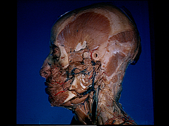
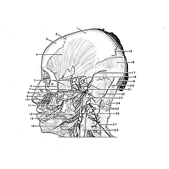
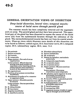
Deep facial dissection, lateral view; temporal muscle; course of facial nerve through parotid gland
Image #49-5
KEYWORDS: Cheek, Connective tissue, Exocrine and endocrine, Face, Muscles and tendons, Peripheral nervous system, Vasculature, Fascia and connective tissue, Overview.
Creative Commons
Stanford holds the copyright to the David L. Bassett anatomical images and has assigned Creative Commons license Attribution-Share Alike 4.0 International to all of the images.

For additional information regarding use and permissions, please contact Dr. Drew Bourn at dbourn@stanford.edu.
|
| ||||||||||||||||||||||||||||||||||||||||||||||||||||||||||||
|
|