Bassett Collection of Stereoscopic Images of Human Anatomy
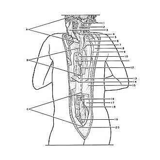
Exploration of the spinal cord and meninges in situ
Orientation view; areas of subsequent close-up views indicated
Image #32-2
KEYWORDS: Bones joints cartilage, Central nervous system, Cervical region, Lumbar region, Meninges, Peripheral nervous system, Sacral region, Thoracic region, Vertebral column, Bones cartilage joints, Cervical vertebrae, Overview.
Creative Commons
Stanford holds the copyright to the David L. Bassett anatomical images and has assigned Creative Commons license Attribution-Share Alike 4.0 International to all of the images.

For additional information regarding use and permissions, please contact Dr. Drew Bourn at dbourn@stanford.edu.

|
| ||||||||||||||||||||||||||||||||||||||||||||

|
|