Bassett Collection of Stereoscopic Images of Human Anatomy
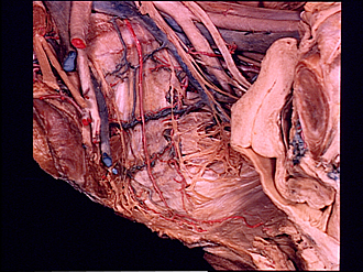
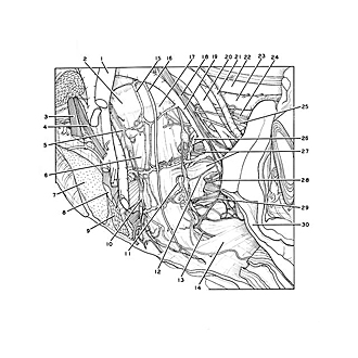
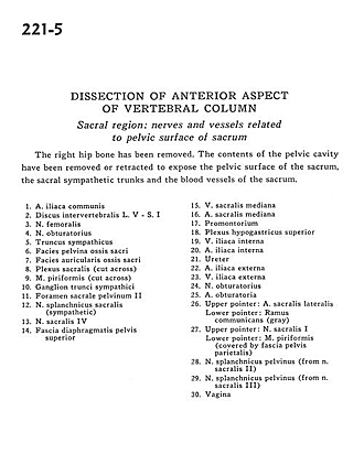
Dissection of anterior aspect of vertebral column
Sacral region.
Image #221-5
KEYWORDS: Sacral region, Vasculature, Vertebral column.
Creative Commons
Stanford holds the copyright to the David L. Bassett anatomical images and has assigned Creative Commons license Attribution-Share Alike 4.0 International to all of the images.

For additional information regarding use and permissions, please contact Dr. Drew Bourn at dbourn@stanford.edu.
|
| ||||||||||||||||||||||||||||||||||||||||||||||||||||||||||||||
|
|